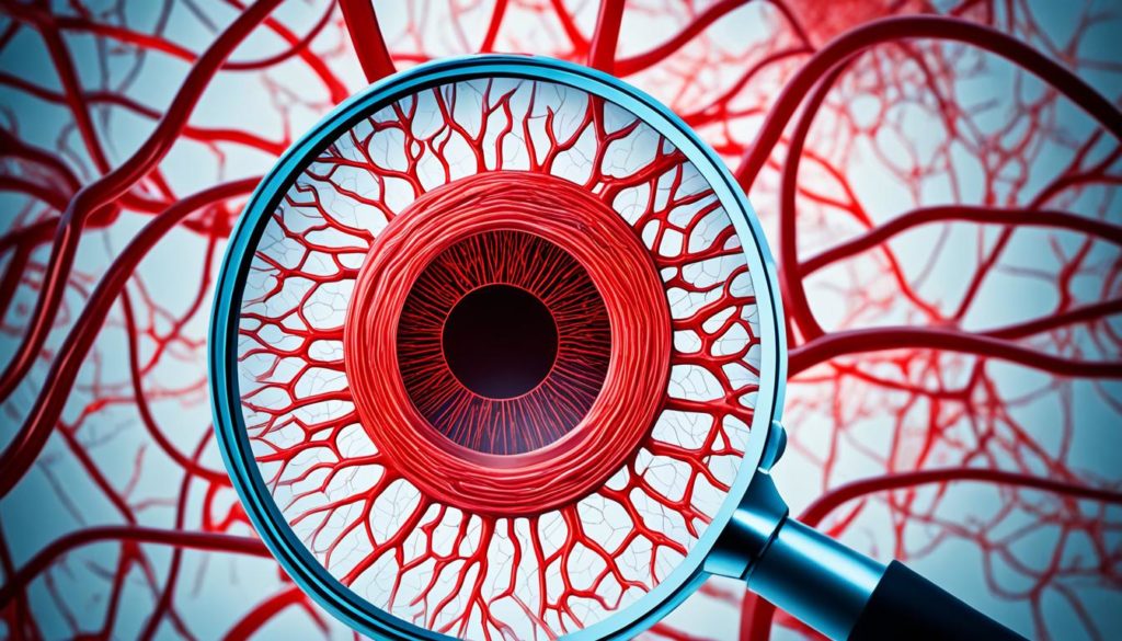
Did you know 5-10% of infants are born with hemangiomas? That makes them a common type of vascular birthmark. It’s vital for both affected individuals and their families to learn about them. Our guide covers the causes, symptoms, how they’re diagnosed, and ways to treat them.
Sourcing our knowledge from the American Academy of Dermatology and top research, readers will understand these benign tumors well. They’ll learn the best ways to manage hemangiomas.
Key Takeaways:
- Hemangiomas affect 5-10% of infants, presenting as common vascular birthmarks.
- Understanding the causes and symptoms of hemangiomas is crucial for early diagnosis and treatment.
- Diagnostic procedures help in identifying the type and severity of hemangiomas.
- Treatment options include medications, laser therapy, and surgery.
- Information from reputable sources like the American Academy of Dermatology is critical for effective management.
What are Hemangiomas?
Hemangiomas are benign tumors that often show up after a baby is born. They’re known for their unique color and feel, made up of extra blood vessels. It’s important to know about hemangiomas to tell them apart from other skin issues.
Definition
Hemangiomas are harmless tumors from endothelial cells. They’re mainly seen in babies. They come in various sizes and places on the body. What sets a hemangioma apart is its blood vessel makeup, making it different from other skin spots.
Common Types of Hemangiomas
There are a few main kinds of hemangiomas. The most common are the strawberry birthmark and the cavernous hemangioma. Each has its own look and growth way.
- Strawberry Birthmark: Known as an infantile hemangioma, this looks like a bright red strawberry surface. These usually stay on the surface but can grow bigger.
- Cavernous Hemangioma: These are deeper than strawberry marks and can look blue or purple. They can disrupt more tissue and sometimes need treatment if they affect important body parts.

Causes of Hemangiomas
The growth of hemangiomas is shaped by both genetic and environmental elements. Recognizing these factors is key for better treatments and prevention.
Genetic Factors
Genetics strongly influence hemangiomas. Ongoing studies show links between genetic mutations and these conditions. Mutations in the VEGF and FGFR genes are important here.
There are clear patterns within families, showing a hereditary aspect. Yet, we need more insights into how genes play their part.
Environmental Influences
Environment also affects hemangiomas. Research points to maternal health, early birth, and low birth weight. There may be risks from certain toxins during pregnancy too.
But it’s still unclear which environmental factors are key. More in-depth studies are needed to pinpoint these triggers.

Scientists are looking into how genes and environment work together. They think environmental factors might trigger hemangiomas in those who are genetically inclined. This could help explain why hemangiomas vary across different groups and situations.
| Genetic Factors | Environmental Influences |
|---|---|
| VEGF gene mutations | Maternal health |
| FGFR gene mutations | Premature birth |
| Familial patterns | Low birth weight |
| Environmental toxins |
Symptoms of Hemangiomas
Hemangiomas are usually known by their looks. They can be tiny spots or large, worrisome growths. Knowing the symptoms is key to early recognition and care.
Visible Signs and Physical Symptoms
The symptoms of hemangiomas vary based on their type and where they are. Common signs include:
- Color: Bright red, blue, or purple hues.
- Shape: Often round or oval.
- Texture: Smooth, raised, or bumpy.
Hemangiomas can also hurt or feel uncomfortable. This is more likely if they’re in a sensitive spot.
Possible Complications
Hemangiomas may cause other health problems, so it’s good to know the risks. Complications range from minor skin issues to serious health concerns. Common problems include:
- Ulceration: Open sores that can get infected.
- Bleeding: This can happen if the hemangioma is hurt or scratched.
- Impaired function: Hemangiomas can affect vital functions if they grow near important parts like the eyes or windpipe.
It’s crucial to tackle these issues quickly. Doing so helps avoid lasting harm or major health problems.
Diagnosing Hemangiomas
The first step in diagnosing hemangiomas is a close look by a dermatologist. They often appear as red, raised marks on the skin. If there’s any doubt, more tests may be needed.
“Medical imaging techniques such as ultrasound and MRI are frequently employed to assess the deeper characteristics and precise size of hemangiomas,” notes the American Academy of Dermatology.
These imaging tools are key to understanding how the lesion affects surrounding tissues. Medical imaging also helps tell hemangiomas apart from other vascular issues. MRI, in particular, gives a clear view of what’s happening inside the body.
Sometimes, if the hemangioma looks unusual or doesn’t get better with treatment, a skin biopsy may be needed. This means taking a tiny piece of the lesion for tests. The pathology report from the biopsy tells us that the growth is harmless.
Let’s compare different ways to diagnose:
| Diagnostic Method | Purpose | Advantages |
|---|---|---|
| Visual Assessment | Initial diagnosis based on appearance | Non-invasive, quick |
| Ultrasound | Assess lesion depth and blood flow | Real-time imaging, cost-effective |
| MRI | Detailed internal structure imaging | High accuracy, differentiates tissue types |
| Skin Biopsy | Histopathological confirmation | Conclusive, identifies malignancy |
Vascular Birthmarks: Strawberry and Cavernous Hemangiomas
Vascular birthmarks, such as strawberry and cavernous hemangiomas, are present at birth or soon after. Knowing the differences between these types is key to choosing the right treatment.
Strawberry Hemangiomas
Strawberry hemangiomas are raised, red birthmarks. They usually show up in the first weeks of life. You’ll often see them on the face, head, back, or chest.
They grow fast and are brightly colored, but most shrink and fade by age five. They rarely need treatment early on unless they affect vital functions. Treatment might include creams or laser therapy when needed.
Cavernous Hemangiomas
Cavernous hemangiomas are deeper and can involve internal organs. They look bluish and are soft to the touch. The treatment depends on their size, place, and complications.
Studies show treatments like interventional radiology or surgery can help. It’s important to keep an eye on them with regular check-ups and imaging.
| Feature | Strawberry Hemangioma | Cavernous Hemangioma |
|---|---|---|
| Appearance | Bright red, raised | Bluish, soft mass |
| Common Locations | Face, scalp, back, chest | Subcutaneous tissues, internal organs |
| Usual Age of Occurrence | Infancy | Any age |
| Treatment Options | Topical medications, laser therapy | Interventional radiology, surgery |
| Prognosis | Often regress by age 5 | Requires monitoring |
Infantile Hemangiomas
Infantile hemangiomas are common benign tumors in newborns. About 4-5% of newborns in the United States have them. These vascular birthmarks often occur in females, premature infants, and low birth weight babies.
Prevalence in Newborns
Newborn skin conditions like hemangiomas usually show up in the first weeks of life. Neonatal medical journals say around 10% of premature babies have them, unlike full-term infants. It’s critical to watch newborn skin closely for early detection and care.
Growth and Regression Phases
The hemangioma growth cycle has two phases: proliferation and involution. Proliferation happens in the first 3-6 months, with noticeable growth. This phase is key for deciding if medical help is needed to avoid problems.
The involution phase lasts for several years, until about age 10. The hemangioma slowly gets smaller during this time, often disappearing. Studies on pediatric dermatology point out the differences in how long this takes and the results, showing the need for careful tracking.
Knowing about the hemangioma growth cycle helps caregivers and medical teams make good choices for treatment. Doctors use set rules to know when to step in, aiming for the best care for these newborns.
Monitoring well and stepping in at the right time during the growth phase can stop problems. It also makes life better for babies with hemangioma growth.
Hemangiomas in Adults
Hemangiomas are mostly seen in infants, but adults can have them too. Adult vascular lesions differ a lot from those in children. This difference makes diagnosis and treatment challenging. Learning about hemangiomas in adulthood is key for proper management.
Adult hemangiomas might stay from childhood or appear for the first time due to certain factors. Research shows these lesions can be on the skin, muscles, bones, or inside the body. Their causes might be genetic or due to the environment.
Treating hemangiomas in adults requires careful monitoring. Sometimes, medication or surgery is needed. If a hemangioma from childhood remains, doctors may suggest various treatments.
| Factors | Infantile Hemangiomas | Hemangiomas in Adults |
|---|---|---|
| Common Locations | Skin, Scalp, Neck | Skin, Muscles, Bones, Internal Organs |
| Treatment Approaches | Observation, Possible Medication | Medication, Surgical Procedures |
| Progression | Typically Regress Naturally | May Persist or Develop Anew |
Understanding adult hemangiomas is getting better. It shows why a detailed clinical check-up and personalized treatment matter. For deeper insights, it’s good to look into varied treatments and ask experts for advice.
Hemangioma Treatment Options
Treating hemangiomas has gotten better with many options available. Each case needs a special plan. Treatments include medicines, lasers, and surgery.
Medications
Doctors often start with medicine to treat hemangiomas. Beta-blockers, like propranolol, work well to make them smaller and less noticeable. If beta-blockers don’t work, corticosteroids are another choice. They can also reduce swelling and help hemangiomas shrink.
Laser Therapy
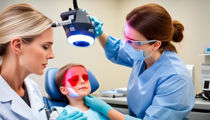
Laser therapy is a skin-friendly choice, great for surface-level hemangiomas. It uses pulsed dye lasers to attack the hemangioma’s blood vessels. This makes them smaller over time. Research backs up laser therapy as safe and helpful for patients of all ages.
Surgery
When other treatments don’t work, removing the hemangioma surgically might be necessary. This is usually for hemangiomas that bother someone a lot or affect how they use a body part. Surgery might be simple or complex, depending on the hemangioma’s size and spot. After surgery, patients need careful follow-up to heal well and avoid big scars.
FAQ
What are hemangiomas?
Hemangiomas are non-cancerous growths of blood vessels that usually appear after a child is born. These growths go through a quick growth stage and then slowly get smaller over time.
What are the common types of hemangiomas?
The main types of hemangiomas are strawberry hemangiomas, which are red and stick out, and cavernous hemangiomas, which are deeper and can look blue or purple.
What causes hemangiomas?
Hemangiomas come from both genes and the environment. While experts know there’s a genetic link, they’re still figuring out the environmental factors.
What are the symptoms of hemangiomas?
Hemangiomas appear as red or purple spots that can be either flat or bumpy. They might cause pain or discomfort. Sometimes, they can lead to issues like sores or affect important body functions if their size and place cause problems.
How are hemangiomas diagnosed?
Doctors identify hemangiomas by looking at them, using imaging tests like ultrasounds or MRIs, and sometimes doing a skin biopsy, especially for unusual cases.
How can hemangiomas in adults appear or persist?
In adults, hemangiomas might stay from childhood or appear new. They can look different and need other ways to manage them compared to hemangiomas in kids.
What are the treatment options for hemangiomas?
Treating hemangiomas can include using medicine like beta-blockers and corticosteroids, laser treatments, or removing them through surgery. The best treatment depends on where the hemangioma is, how big it is, and if it’s causing problems.
What are strawberry hemangiomas?
Strawberry hemangiomas are vascular birthmarks that are bright red and raised. They mostly occur in babies and grow fast at first, then shrink, often going away by age 10.
What are cavernous hemangiomas?
Cavernous hemangiomas are deeper and can be blue or purple. They’re made of larger blood vessels than strawberry hemangiomas and can affect nearby areas depending on their size and spot.
What is the prevalence of infantile hemangiomas?
Infantile hemangiomas are fairly common, with about 5-10% of infants having them. They typically show up a few weeks after birth and grow, then shrink over years.
What should I expect during the growth and regression phases of infantile hemangiomas?
In the growth phase, infantile hemangiomas can quickly get bigger and more noticeable. After that, they slowly become smaller and fade during the regression phase. By around ages 7 to 10, most hemangiomas decrease a lot in size.


