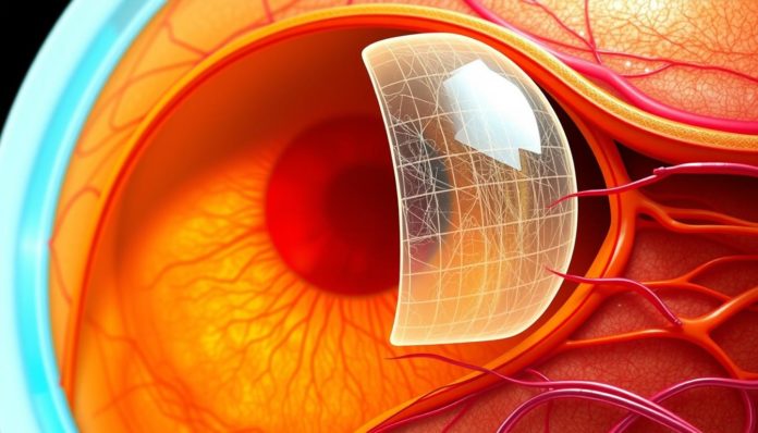
An Epiretinal Membrane (ERM) is a concern for people facing this eye issue. This layer of tissue can cause something called macular pucker. It’s key to know about ERM treatment options. Learning about this helps you understand why seeing a retinal specialist is so important.
We want to help you understand ERM well. It’s not just about the facts. It’s about improving life quality for those with ERM. Let’s explore how knowledge leads to better actions and relief.
What Is an Epiretinal Membrane?
An epiretinal membrane (ERM) is a thin layer on the retina’s surface. It’s found over the macula, vital for clear central vision. Known as macular pucker, it can distort vision by wrinkling the retina. Learning about the Epiretinal Membrane definition, how it affects sight, and how it differs from other macula issues is key to handling this eye problem.

The Basics of Epiretinal Membrane
Epiretinal membranes are mostly due to changes in the eye’s vitreous. This gel inside the eye shrinks and may pull away from the retina as we age. This can make scar tissue form, causing the macula to wrinkle or pucker. Such wrinkling can make straight lines look wavy and text seem blurry. Watching this condition through regular check-ups and OCT scans helps in knowing if it gets worse or stays the same.
Distinguishing Between Epiretinal Membrane and Other Eye Conditions
Even though epiretinal membranes can lead to vision problems like other eye diseases, there are clear differences. For instance, macular degeneration happens when the macula’s surrounding tissues age and thin, needing a different treatment. On the other hand, ERMs are physically caused by the membrane’s presence. Sometimes, surgery can remove the membrane to either improve or keep vision stable.
The Signs and Symptoms of ERM
Epiretinal membranes (ERMs) present unique challenges, mainly showing through ERM symptoms like vision changes. People may notice their vision distorting, becoming less sharp, and having trouble with small details. It’s key to recognize these signs early and seek help from a retinal specialist.

One major sign of ERM is vision distortion. Straight lines might look bent or wrong. This can make daily tasks difficult, such as reading, driving, or recognizing people. Also, vision may get blurry or seem like a gray curtain is moving across what you see. These issues can get worse without treatment.
- Persistent blurriness in central vision
- Difficulty seeing fine details, such as small print in books or delicate handwork
- Distorted perception of straight lines, which may appear wavy or curved
- Double vision when looking with one eye (monocular diplopia)
Seeing any ERM signs means you should talk to a retinal specialist. Catching and managing ERM early can save your sight and may avoid serious procedures. A specialist can provide a full look at your condition and talk about the best treatments for you.
Not seeking help early can make symptoms worse, even leading to lasting vision loss. It’s vital to get checked early to spot ERMs and rule out other eye problems.
Epiretinal Membrane: A Closer Look at the Causes
Understanding why epiretinal membrane (ERM) happens is key for everyone involved. We’re going to explore the main causes of ERM. Age and genetics are big factors in this eye problem.
Common Risk Factors for Epiretinal Membrane Development
There are many risk factors for ERM, but some stand out more. Things like eye surgery, diabetes, retinal tears, and inflammation contribute. Knowing these risks can help catch and treat ERM early.
How Age and Genetics Influence Epiretinal Membrane
Getting older is a big risk for ERM. As we age, changes in our eyes make ERM more likely. Genetics matter too. Some genes make you more likely to get ERM. If ERM runs in your family, your risk goes up.
This look into ERM causes shows who’s most at risk because of age and genetics. It highlights why knowing your risk is crucial for preventing and treating ERM.
| Age Group | Incidence Rate of ERM | Genetic Influence (High/Low) |
|---|---|---|
| 50-60 years | 2% | Low |
| 61-70 years | 5% | Moderate |
| 71 years and above | 9.7% | High |
Exploring Diagnostic Procedures for Epiretinal Membrane
An effective ERM diagnosis depends on the retinal specialist’s skill and diagnostic tools. It’s key for deciding on eye surgery or other treatments.
Optical Coherence Tomography (OCT) leads in diagnosing epiretinal membranes. This test is safe and gives detailed pictures of the retina. These images show abnormal membranes or distortions.
- OCT is key in finding ERM’s severity and details.
- Its clear images aid in planning surgery, if needed.
Fundus photography also plays a crucial role by capturing the retina in detail. It tracks the disease over time. Combined with OCT, it offers a full view of the retinal surface.
Spotting epiretinal membranes early through detailed imaging is vital for managing and treating them successfully.
Sometimes, when symptoms and tests suggest ERM, but images don’t confirm, more tests are needed. A retinal specialist might use Fluorescein Angiography (FA). FA shows blood flow and the health of retina’s vessels, often impacted by ERM.
In the end, these diagnostic tools confirm ERM’s presence. They guide in creating a tailored treatment plan. This might include eye surgery, medications, or monitoring the condition.
Epiretinal Membrane and Its Connection with Macular Pucker
Learning about the ERM connection with macular pucker is vital. It helps us understand their relationship and how they differ. This knowledge is key for better diagnosis and treatment.
Understanding the Relationship Between ERM and Macular Pucker
ERM, also known as epiretinal membrane, often leads to a condition called macular pucker. This happens when a thin layer forms on the retina’s surface. Specifically, it affects the macula, which is crucial for clear vision.
Identifying the Differences: ERM vs. Macular Pucker
The term “macular pucker” refers to the puckering seen on the macula. Professionals use it to describe visible, symptomatic changes. “ERM”, however, describes thin membranes that could appear anywhere on the retina’s inner layer. It’s not just limited to the macula.
This understanding clarifies the differences between macular pucker and other conditions. It makes sure patients get the right treatment based on accurate diagnoses.
The Role of a Retinal Specialist in ERM Treatment
When diagnosed with an Epiretinal Membrane (ERM), patients seek help from a retinal specialist. Their expertise is key in diagnosing and managing ERM. They create personalized treatment plans to meet each patient’s unique needs.
Retinal specialists are well-trained in eye diseases. They know the retina and vitreous body well. This helps them in identifying ERMs using advanced imaging. They then explain the best treatment options to improve or save vision.
- Initial Consultation and Diagnostic Imaging
- Customized Treatment Plan Development
- Regular Monitoring and Adjustments to Treatment
- Long-term Rehabilitation Guidance
Having a retinal specialist involved in ERM therapy is crucial. They keep improving their methods with new research and clinical trials. Their skill in using new techniques is vital for effective ERM care.
Patient education is also important. Retinal specialists make sure patients understand their condition and treatment. This empowers patients, making them active in their recovery journey.
The main aim of a retinal specialist is to enhance the patient’s life. They do this by providing advanced, precise, and caring treatment for ERM.
Epiretinal Membrane Treatment Options
Patients with an epiretinal membrane (ERM) have several treatment choices. They can choose between non-surgical methods or eye surgery. This depends on how severe the condition is and their symptoms.
Non-Surgical Management of Epiretinal Membrane
Non-surgical options often help patients manage their symptoms well. Observation is one strategy, especially for mild symptoms or when the membrane doesn’t majorly affect daily life. Medication can also help with the symptoms linked to the membrane.
When is Surgery Recommended for Epiretinal Membrane?
Surgery becomes an option when the ERM seriously affects vision or symptoms get worse. The main surgery for this is a Vitrectomy. It removes the eye’s vitreous gel for better access to the retina. The surgeon can then remove the membrane, which helps ease tension and improve vision.
Vitrectomy is key for those with severe ERM issues. It offers a chance to get better vision and quality of life back.
- Observation and regular monitoring for mild cases
- Medication to alleviate associated symptoms
- Vitrectomy for significant vision impairment
Knowing all ERM treatment options, including non-surgical and surgical, is vital. It helps patients and caregivers effectively manage this eye condition.
Understanding the Vitrectomy Procedure for ERM Surgery
The vitrectomy procedure is vital for treating epiretinal membranes (ERM). It involves detailed methods carried out by a retinal specialist. This surgery is key to improving how well you see and fixing vision problems caused by ERM. This section will explore the main stages of vitrectomy, helping patients know what happens during ERM surgery.
First, the retinal specialist checks the patient’s eye to see if a vitrectomy is needed. The doctor does thorough eye exams and plans the surgery based on the patient’s specific ERM details.
- Preoperative assessment and planning
- Anesthesia administration to ensure comfort
- Removal of vitreous gel to grant access to the retina
- Peeling of the epiretinal membrane
- Replacement of vitreous with a saline solution to maintain eye pressure
- Postoperative care to monitor recovery and mitigate risks
The vitrectomy is performed using precise tools through tiny incisions. This minimizes harm to the eye. The success of ERM surgery largely relies on the retinal specialist’s skill and the ERM’s complexity.
After surgery, patients may see clearer and have less visual distortion. This depends on their specific case and the ERM’s severity before the operation. Regular visits with the retinal specialist are important for healing and handling any complications after surgery.
Knowing about the vitrectomy process helps patients make smart choices about their ERM surgery. They can have realistic hopes for better vision. With a careful retinal specialist, the path to improved sight can be well navigated.
Recovery and Prognosis After ERM Surgery
After ERM surgery, patients often wonder about their recovery and how their vision might improve. We will explore the common recovery timeline and long-term vision outcomes. This procedure has promising results for many.
What to Expect During the Recovery Period
The recovery from ERM surgery involves a few key steps. At first, patients might have mild discomfort and blurry vision. These symptoms generally get better as the eye heals. It’s critical to stick to your surgeon’s care plan, including eye drops for preventing infection and reducing swelling. Not doing heavy lifting and keeping your head in the right position are also important.
Make sure you don’t rub or press on your treated eye. Always go to your follow-up checks with your eye doctor. And watch out for signs that something’s wrong, like more pain or vision getting worse suddenly.
Long-Term Outcomes of ERM Surgery
Most people see a big improvement in their vision after ERM surgery. How well the surgery works mostly depends on how serious the membrane was and the eye’s health before surgery. A lot of patients find their vision gets better or stabilizes a few months after the surgery.
To give you a better idea, here’s a chart with common results seen after ERM surgery:
| Time Post-Surgery | Vision Improvement | Additional Notes |
|---|---|---|
| 1 Month | Mild to Moderate | Continued use of medicated eye drops |
| 3-6 Months | Moderate to Significant | Possible adjustment of glasses prescription |
| 1 Year | Most noticeable improvement | Stable vision with minimal fluctuations |
Knowing about the recovery and long-term chances of better vision can help patients have realistic hopes. It leads to an easier recovery and a better final result. Regular talks with your eye doctor will help catch any issues early. They make sure your journey to better vision improvement is successful.
Potential Complications and Risks of ERM Surgery
ERM surgery is usually safe but carries some risks and complications. Knowing these helps patients make informed choices. They can spot problems early after surgery. Safety in eye surgery is key to reducing these risks.
Some common complications could happen during or after ERM surgery. We’ll talk about these issues and how to handle or avoid them.
- Infection: They’re rare but can happen. They’re treated with antibiotics.
- Hemorrhaging: There might be minor bleeding in the eye during surgery.
- Retinal Detachment: This serious issue may need more surgery to fix.
- Cataract Development: It’s common after surgery and might require more surgery later.
To improve eye surgery safety, prevention is important. This includes careful checks before surgery and following post-surgery care closely. Patients should listen to their doctor’s advice to lower risks.
| Complication | Incidence Rate | Preventive Measures |
|---|---|---|
| Infection | Less than 1% | Sterile surgical environment, post-op antibiotics |
| Hemorrhaging | 1-2% | Precise surgical techniques, immediate post-op care |
| Retinal Detachment | About 3% | Timely diagnosis and treatment, regular follow-ups |
| Cataract Development | Over 20% within two years | Monitoring and early treatment as needed |
In conclusion, ERM surgery’s potential complications may seem scary, but medical advancements and safety steps greatly lower these risks. Knowing about these issues, along with expert help, ensures ERM surgery’s safety and success.
Advancements in ERM Treatment and Eye Surgery
The way we treat epiretinal membranes (ERM) has changed a lot, thanks to new ERM treatment advancements. These improvements have made patient results better and allowed for more types of procedures. As we keep researching, the ways we do eye surgery are setting new standards.
Innovations in Vitrectomy Techniques and Equipment
Vitrectomy tools have gotten a big upgrade recently, showing off major eye surgery innovations. Now, we have better micro-incision tools. They help patients recover faster and let doctors take out membranes more precisely. Also, we now have better imaging tech during surgeries. This lets doctors see the retina clearer, making surgeries safer and more accurate.
Future Directions in ERM Treatment Research
Looking forward, we want to find ways to treat ERM without surgery. Researchers are exploring drug treatments that might make surgery unnecessary. They’re also looking into how our genes affect ERM. This could lead to personalized treatments, aiming to improve the health and vision of our eyes the most.
Living with Vision Distortion Caused by Epiretinal Membrane
Having an Epiretinal Membrane (ERM) means facing big changes. Vision distortion makes simple tasks hard. Reading, recognizing faces, and walking in crowds can become tough. How much this affects someone’s life depends on the severity of the distortion. But, people with ERM find that changing daily routines and using special visual aids helps a lot.
Making some lifestyle changes helps manage the condition better. More light can make seeing clearer. Using materials that have high contrast and big print also helps ease eye strain. Plus, gadgets with options to enlarge text or talk back can make a big difference. These tools help people with ERM stay independent and enjoy life more.
Having regular check-ups with a retina specialist is key to managing ERM. Eye doctors give advice on new treatments that might improve vision. Also, joining support groups to meet others with similar experiences is helpful. These groups provide both emotional support and practical tips. Though ERM means adjusting, with the right management, people can still lead active, happy lives.
FAQ
What exactly is an Epiretinal Membrane?
An Epiretinal Membrane (ERM) is a thin tissue over the macula. The macula is for sharp vision. ERM can distort vision and cause blurring.
How can I tell the difference between Epiretinal Membrane and Macular Degeneration?
ERM adds a layer on the retina. Macular Degeneration causes the macula to weaken. An eye expert can tell them apart with exams and OCT scans.
What are common symptoms of Epiretinal Membrane?
ERM symptoms include blurry and distorted vision. You might see straight lines as wavy. It can make detailed work hard. Some may see double in one eye.
What causes an Epiretinal Membrane to develop?
ERMs’ causes are often unknown. They’re common after 50. Causes include retinal tears or surgery. Sometimes, genes play a part.
What’s the connection between an Epiretinal Membrane and Macular Pucker?
Epiretinal Membrane and macular pucker mean the same thing. “Macular pucker” describes how ERM makes the retina wrinkle.
What role does a retinal specialist play in ERM treatment?
A retinal specialist diagnoses ERM and its effect on sight. They recommend treatment, which could be observation or surgery.
What treatment options are available for Epiretinal Membrane?
For ERM, treatment may just be watching the condition. Or it may mean surgery. A common surgery removes the ERM to improve vision.
What can I expect during the recovery period after ERM surgery?
Recovery from ERM surgery can take weeks to months. You might need to keep your head in a special position. Vision usually gets better as healing happens.
Are there any risks involved with ERM surgery?
ERM surgery, like any surgery, has risks. These include infection and bleeding. Retinal detachment is another risk. But these problems are rare. The surgery is safe overall.
Have there been any recent advancements in ERM treatment?
Yes, surgery for ERM has gotten safer with new tools. These include better microscopes. There’s research into less invasive treatments and better care after surgery.
How can I manage daily life with vision distortion from ERM?
People with ERM use visual aids and change their surroundings. Increasing light and contrast helps. Regular eye care is also key to living well with vision distortion.


