Keeping your eyes healthy is all about precision. Fluorescein Angiography is a top tool for eye doctors. It shines a light on the retina’s blood system. This method finds retinal problems early, which can stop serious eye diseases.
This ophthalmology test shows blood movement in the retina. It can catch signs of trouble early. Doctors worldwide use it to keep eyes healthy. It’s a key tool for them, showing its value in vision care.
Understanding Fluorescein Angiography
Fluorescein angiography is key in diagnosing and managing retinal diseases. This diagnostic procedure uses a dye injection test. It creates clear images of the eye. These images help us understand the retina’s blood vessels better.
What is Fluorescein Angiography?
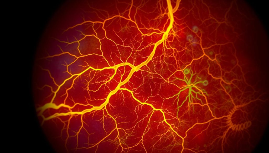
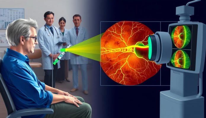
Fluorescein angiography is a special way to see the retina’s blood flow. It uses a fluorescent dye injected into the bloodstream. As the dye moves, a special camera takes pictures of the retina. These photos show if there are any problems with the blood vessels.
The History and Development of Fluorescein Angiography
Fluorescein angiography has evolved from a new idea to a vital diagnostic procedure. It started in the 1960s and changed how we find retinal diseases. With better technology, this test has become essential in eye care today.

Why Fluorescein Angiography is a Crucial Diagnostic Tool
Fluorescein angiography is a key diagnostic method. It excels in showing retinal blood vessels in detail. This approach is vital for spotting and helping with different retinal diseases, like the tricky macular degeneration.
This test is great for looking at blood flow in the retina. It lets eye experts check the health of retinal blood vessels. Changes in these vessels often point to early retinal problems. Finding these issues early with angiography means treatments might work better, maybe even stopping diseases.

In dealing with macular degeneration, which can cause loss of vision, this tool is crucial. It spots unusual blood vessel activities under the retina, key in identifying wet macular degeneration. This helps in quick and right diagnoses, leading to specific treatments like VEGF inhibitor injections.
- Enhanced visualization of the retinal blood vessels
- Early detection of retinal diseases like macular degeneration
- Guidance for effective treatment plans
Thus, the ongoing use and improvement of fluorescein angiography are key. They’ll help us better understand and treat retinal diseases. This means patients all over the world could enjoy better vision outcomes.
The Fluorescein Dye Injection Test Explained
The fluorescein dye injection test is key in spotting retinal diseases. It helps keep eyes healthy. We’ll cover the steps involved. This ensures patients know what happens during the test.
Preparing for the Test
Getting ready for the test starts with checking the patient’s medical history. This is to make sure the dye is safe for them. Patients should not wear contact lenses. They also need someone to drive them home because their vision might blur a bit.
The Injection Process
The injection is quick and done in a clean way. A special dye that helps see the retina is used. It goes into a vein in the arm. Then, it travels to the eye, showing the blood vessels in the retina.
What Happens After the Dye is Administered?
Right after the dye goes in, things move fast. Patients get a few rapid eye photos taken. These photos show how the dye moves in the eye. They help find any blockages or leaks, giving doctors the info they need.
Fluorescein Angiography in Action: Capturing Retinal Images
Fluorescein angiography is an advanced way to look at retinal images. It helps eye doctors see important details, key in treating eye issues.
The Role of Digital Imaging in Fluorescein Angiography
Digital tech has changed fluorescein angiography. It offers clear images of the retina. This is crucial for checking the eye’s health.
It also spots any strange spots or lesions. Digital images are quick to get. This means patients spend less time being checked.
Interpreting the Results
After taking images of the retina, the next step is to figure them out. Experts look for signs like leaks, blockages, or problem vessels. These are hints of issues like diabetic retinopathy or macular degeneration.
Reading these images right is vital. It helps in making a plan that suits the patient’s needs.
| Indicator | Normal Condition | Abnormal Condition |
|---|---|---|
| Vessel Appearance | Uniform and unobstructed | Irregular width, leakage |
| Fluorescein Flow | Consistent throughout the retina | Delayed or sparse in regions |
| Leakage Areas | None | Visible near or around the vessels |
By using advanced digital methods and careful study, fluorescein angiography gets better at imaging the retina. It’s a key tool for eye doctors.
Diagnosing with Fluorescein Angiography
In the world of eye health, spotting and diagnosing retinal diseases early is key. Fluorescein angiography is a top method for checking retinal blood vessels. It gives doctors important images that help spot issues and plan the right treatment.
Fluorescein angiography’s main job is finding problems in the retinal blood vessels. These issues could signal serious retinal diseases. A special dye is used for this. It shows how blood flows in the retina. This lets doctors see any blockages or leaks in real time.
Here’s a list of conditions fluorescein angiography is especially good for:
- Diabetic retinopathy: This disease hurts the retinal blood vessels in people with diabetes, causing vision problems. Spotting it early with this technique can stop worse issues.
- Age-related macular degeneration (AMD): For AMD, fluorescein angiography spots leaking vessels under the retina. This info is useful for treatments like laser therapy.
- Vascular occlusions: It’s key for seeing retinal vein occlusion, where a vein blockage can cause sudden blindness.
Fluorescein angiography isn’t just for finding diseases. It also checks how well treatments are working and how diseases progress. The clear images from this method allow doctors to care for their patients better.
What to Expect During a Fluorescein Angiography Examination
Getting a fluorescein angiography is a key part of keeping your eye health in check. It’s really helpful if you’ve had eye problems that need a close look. Let’s go through what happens from start to finish.
Before the Procedure
Before the test, it’s crucial to talk about any medical concerns with your doctor. This includes allergies. Make sure not to wear contact lenses. You’ll also need someone to drive you home since the dye injection test might blur your vision for a bit.
During the Procedure
The main event is the dye injection test. A dye called fluorescein will be put into your veins. It helps show the blood vessels in your eye’s back part. The injection might give a quick pinch, but it’s over fast. After that, pictures are taken as the dye moves around. This lets the doctor see how blood is flowing in your retinal vessels.
After the Procedure
After the angiography, you might see your skin or pee turn a bit yellow. This is normal and goes away after a few hours. It’s best to take it easy and rest your eyes. Avoid hard activities for the day. Your eye doctor will tell you when to come back to talk about what they found.
Knowing what to expect before, during, and after a fluorescein angiography can ease your worries. It helps you get ready for an important eye examination. This test plays a big part in maintaining your eye health for the long run.
Comparing Fluorescein Angiography with Other Retinal Imaging Techniques
Medical technology is growing fast, making the choice of diagnostic tests for the eye’s retina very important. Fluorescein angiography has been vital in eye care, showing the retina’s blood vessels in detail. But now, there are more options like Optical Coherence Tomography (OCT) helping doctors meet their patients’ specific needs.
OCT vs. Fluorescein Angiography
Optical Coherence Tomography (OCT) and fluorescein angiography are key for eye disease care but are used differently. OCT uses light to create detailed retina images, crucial for spotting conditions such as macular holes. Fluorescein angiography, however, uses a dye to make blood vessels visible. This is vital for treating vascular issues like diabetic retinopathy.
Benefits over Traditional Imaging Methods
Fluorescein angiography stands out from older imaging methods. It captures blood flow in real-time, allowing for accurate diagnoses and better treatment plans. This real-time capture of blood circulation is something static imaging can’t offer, proving its worth in specific diagnostic needs.
Knowing when and why to use each imaging tool is key for top-notch eye care. Both OCT and fluorescein angiography serve important roles in ophthalmology. They show how different imaging types can together improve healthcare for the eyes.
Potential Risks and Side Effects of Fluorescein Angiography
Fluorescein Angiography is a key diagnostic procedure for eye health. It’s important to know the risks and side effects. This part will cover what to expect during and after the dye injection test.
Side effects are often mild and go away quickly. Still, knowing about potential complications is vital. It helps patients get ready and feel more at ease.
- Nausea and Vomiting: A common reaction occurring shortly after the dye injection, usually subsiding within minutes.
- Skin Discoloration: The dye may cause temporary yellow-orange discoloration of the skin and urine.
- Allergic Reactions: While rare, some individuals may exhibit allergic reactions ranging from mild itching to severe anaphylaxis.
- Eye Irritation: Patients might experience discomfort or mild irritation in the eye, which typically diminishes soon after the test.
By understanding these risks, patients can talk more easily with their doctors. This helps make choices about the retinal imaging with a clear mind.
This detailed guide aims to educate and get patients ready. Fluorescein angiography is critical for retinal imaging. It’s a powerful way to find and treat many conditions.
Fluorescein Angiography for Diagnosing Retinal Diseases
Fluorescein angiography is crucial in eye care, especially for retinal problems like macular degeneration and diabetic retinopathy. It uses a special dye to show blood flow in the retina. This is key for accurate diagnosis and choosing the right treatment.
Macular Degeneration and Fluorescein Angiography
For those with macular degeneration, this test is vital. It reveals the condition of blood vessels in the eye. Spotting problem areas early can prevent significant vision loss.
Diabetic Retinopathy Detection
It’s also essential for finding diabetic retinopathy soon. Early detection means treatments can start sooner, slowing the disease down. It’s great at showing where blood vessels are leaking, which is common in severe cases.
- Detailed visualization of retinal conditions
- Early detection of retinal diseases
- Precise assessment for targeted treatments
Fluorescein angiography doesn’t just find retinal diseases. It also checks how well treatments are working. This makes it a key part of treating eye conditions like macular degeneration.
Advancements in Fluorescein Angiography Techniques
The field of ophthalmology is rapidly advancing. We see significant improvements in fluorescein angiography, essential for diagnosing and managing eye conditions. These improvements come from better dye injection methods and retinal imaging equipment. They mark a new phase in diagnostic accuracy and better safety for patients.
Innovations in Retinal Dye Technology
Retinal dye technology has seen major upgrades. These include more reactive dyes for clearer, more detailed retinal images. This makes it easier for doctors to spot problems accurately. Also, the new dyes reduce the risk of allergic reactions, making the test safer.
The Future of Retinal Imaging
The future of retinal imaging looks very bright, with ongoing innovations. Expect better resolution and deeper imaging. Artificial intelligence and machine learning are set to change fluorescein angiography. They will make analyzing easier and improve early detection of eye conditions.
Below is a table highlighting the differences between traditional and advanced fluorescein angiography techniques:
| Aspect | Traditional Technique | Advanced Technique |
|---|---|---|
| Image Clarity | Standard Definition | High Definition |
| Safety | Higher risk of reactions | Reduced risk with improved dyes |
| Diagnostic Accuracy | General abnormalities detected | Precise abnormalities detected |
| Technological Integration | Minimal | AI and Machine Learning enabled |
How Fluorescein Angiography Enhances Overall Eye Health
Fluorescein angiography is a key diagnostic procedure in retinal imaging. It greatly benefits eye health. This method helps detect problems early and diagnose them correctly. This way, it’s easier to manage eye conditions and improve patients’ lives.
It uses special dye and a camera to get clear pictures of the retina’s blood vessels. These pictures help find blockages, leaks, or other issues. Catching these signs early helps stop diseases from getting worse and saves vision.
This retinal imaging technique is known for its precision and quickness. It’s a top choice for doctors. The images provide important information. This information helps create personalized treatments for each patient. This makes the treatments more effective.
- Early detection of retinal diseases
- Accurate mapping of retinal blood vessels
- Precise assessment of retinal health
- Guided treatment planning and monitoring
Also, fluorescein angiography is non-invasive. This makes it comfortable and safe for patients. It’s very helpful for regular eye health checks. For those worried about their retinal health, this technique can offer reassurance. It provides detailed and trustworthy health assessments.
When to Consider Fluorescein Angiography
Fluorescein angiography is key in finding and managing retinal diseases. It involves injecting a dye to test for complications. Patients with symptoms like sudden vision changes, floaters, or eye exam irregularities might need it.
Doctors look at symptoms and medical history to decide on this test. Diabetics with signs of diabetic retinopathy, a condition that harms the retina’s blood vessels, often need it. People facing age-related macular degeneration or vision issues might also be candidates.
The decision for this test comes after a careful check by a retina expert. Fluorescein angiography shows important details about the retina’s health. It helps doctors plan treatments early. This test is vital for understanding the eye’s veins and keeping eyes healthy.
FAQ
What exactly is Fluorescein Angiography?
Fluorescein Angiography is a medical test. It uses a special dye and camera to view blood vessels in the retina. It helps diagnose retinal diseases.
How has Fluorescein Angiography changed over time?
At first, images were not very clear. Now, with better cameras and dyes, the pictures are clearer. This makes diagnosis more accurate.
Why is Fluorescein Angiography considered a crucial diagnostic tool for eye health?
It’s vital for seeing retinal blood flow. Doctors can spot diseases early, like macular degeneration or diabetic retinopathy.
What should I expect when preparing for a Fluorescein Angiography test?
Your pupils will be dilated. You should not drive afterwards. The dye may also change the color of your skin and urine for a short time.
What happens after the Fluorescein dye is injected?
The dye moves through the blood to your eyes. Then, the doctor takes photos of your retina. This shows any problems.
Can Fluorescein Angiography results be immediately interpreted?
Yes, the doctor can often see the results fast. This lets you start treatment quickly if needed.
What kinds of retinal diseases can be diagnosed with Fluorescein Angiography?
It can spot many issues, like macular degeneration and diabetic retinopathy. It checks the health of eye blood vessels.
Is the Fluorescein dye injection painful?
Most people only feel a small pinch. It’s not usually painful.
How does Fluorescein Angiography compare to OCT?
OCT gives detailed pictures of the retina. But, Fluorescein Angiography shows blood flow and vessels. It’s better for some diagnoses.
Are there any risks or side effects associated with Fluorescein Angiography?
Some might feel sick or vomit. Allergic reactions can happen but are rare. Quick medical help is important for serious reactions.
How does Fluorescein Angiography contribute to overall eye health?
It helps find retinal problems early. This means better treatment and eye health later on.
When should I consider getting a Fluorescein Angiography test?
If you notice vision changes or spots, or if you have diabetes. Your eye doctor might suggest it.


