Eye health is key to experiencing the world around us. Conditions like Retinal Detachment are serious yet can sneak up on us, deeply affecting our sight. Knowing the signs of Retinal Detachment and its risks is crucial. It is vital for protecting your vision and avoiding lasting harm.
Our future articles will carefully lead you through Retinal Detachment’s aspects. From first warning signs to understanding treatment choices. We aim to arm you with important knowledge. This can help catch problems early and act fast, ensuring your vision stays sharp and your eyes stay healthy.
What is Retinal Detachment?
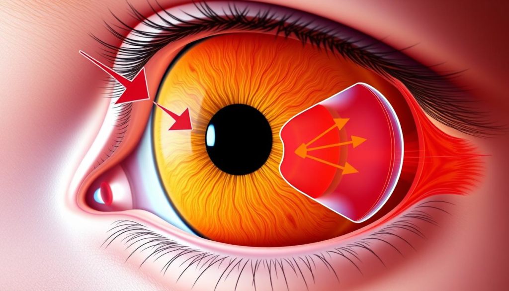
Retinal detachment is when the retina, a thin tissue at the eye’s back, comes off its support layers. This stops the retina from working with light, which is key for seeing. If not fixed quickly, it can cause lasting vision loss.

The Basics of a Detached Retina
The retina is like a camera’s film; it takes in light, changes it to signals, and sends these to the brain. Keeping the retina in place is vital. With a detached retina, it moves from its normal spot, leading to symptoms of retinal detachment like blurry vision or seeing shadows.
How a Retina Becomes Detached: The Medical Process
Detachment usually comes from the retinal layer separating. This can happen because of injury, diabetes eye issues, or getting older. If the retina detaches, it loses oxygen and food. Without putting it back, the retina’s cells start to die, which means vision might not come back.
Knowing the signs of retinal detachment and what causes retinal layer separation is crucial. It means you can get help fast, which greatly improves the chance of saving your sight.
Retinal Detachment Causes and Who is at Risk
To prevent and treat retinal detachment, it’s vital to know its causes and risk factors. We’ll discuss what leads to retinal detachment, who is most at risk, and conditions that increase the chances of getting it.
Underlying Health Conditions Leading to Retinal Detachment
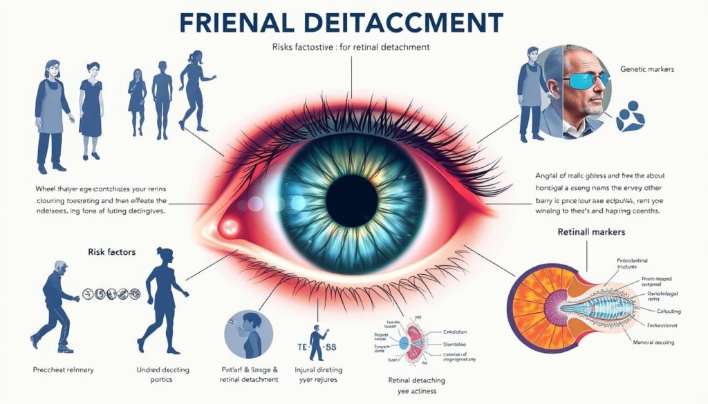
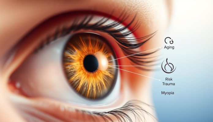
Certain health issues can make retinal detachment more likely. Severe myopia, diabetic retinopathy, and eye injuries are to blame. A family history of retinal detachment also increases the risk, suggesting genetics play a role.
Age-Related Risks for Retinal Detachment
Getting older increases the risk of retinal detachment. The eye’s gel-like substance can shrink and pull away from the retina. This creates a condition called PVD, leading to tears and possibly detachment, especially after 60.
Lifestyle Factors Influencing Detachment Risks
Lifestyle and environment also affect retinal detachment risk. High-impact sports and heavy lifting can harm the eye. Not wearing eye protection increases this risk.
| Risk Factor | Impact | Preventive Measures |
|---|---|---|
| High Myopia | Increases tension on retina | Regular eye exams to monitor changes |
| Ageing | Vitreous degeneration | Monitoring for PVD symptoms |
| Sports/Physical Activities | Potential for trauma | Use of protective eyewear |
| Diabetic Retinopathy | Weakens retina | Control of blood sugar levels |

Retinal Detachment: Signs and Symptoms to Watch Out For
It’s vital to catch the symptoms of retinal detachment early. This condition happens when the retina pulls away from its support tissue. Without quick treatment, it can cause lasting vision loss. Knowing the main symptoms and changes in vision can greatly help.
The first sign often noticed is the sudden appearance of eye floaters. These small, shadowy shapes float through your vision. A sudden increase in floaters is a warning sign that shouldn’t be ignored.
Seeing flashing lights or sparks can also alarm you. This may mean the retina is being pulled on. If it continues, the retina could detach.
A shadow or curtain over your vision is another significant symptom. It generally happens quickly. It’s a sign that the retina might be coming loose.
These visual changes are key warnings. The table below shows common symptoms of this condition. It helps you know when there might be a problem with the retina:
| Symptom | Description | Urgency Level |
|---|---|---|
| Eye Floaters | Increased number of small, dark shapes moving in the field of vision. | Monitor and consult |
| Flashes of Light | Brief, sporadic flashes of light similar to lightning streaks in vision, especially in dim light. | Immediate consultation |
| Darkening Vision | A shadow or dark curtain obstructing part of the visual field, progressing towards central vision. | Emergency |
Noticing these symptoms of retinal detachment early and getting fast medical help can lead to a better outcome. This shows why regular eye exams are important, especially if any symptoms show up.
Stages of Retinal Detachment and Progression
It’s vital to know how retinal detachment progresses. It usually starts with a retinal tear. If ignored, this can lead to part or all of the retina detaching. This puts your sight at risk.
The first signs might be sudden light flashes or an uptick in floaters in your view. These signs typically mean there’s a retinal tear. The retina starts to separate from its support layer. Without quick treatment, the tear may grow.
- Partial Detachment: At this stage, you might notice a shadow or a curtain effect in part of your view. This indicates the retina detaching more and more.
- Complete Detachment: Without stopping the damage, the detachment can become whole. Then, loss of vision gets worse. It could lead to lasting blindness if not treated right away.
A tear can turn into a full detachment in hours to weeks. So, catching it early and getting treated is key to saving your sight. Below, you’ll find more about symptoms and risks at each stage:
| Stage | Symptoms | Risk of Permanent Vision Loss |
|---|---|---|
| Retinal Tear | Flashes of light, many new floaters | Low, if treated quickly |
| Partial Detachment | Visual shadow or curtain effect | Medium, grows over time |
| Complete Detachment | Big loss of vision, darkness | High, needs fast treatment |
Catching and treating this early is super important. Make sure to get your eyes checked often. Especially if you notice any early warning signs. Acting fast at the first hint of a retinal tear can make a huge difference. It might even save your sight.
Seeking Medical Attention: When to See a Doctor
Knowing when to get help for eye issues is key. Early discovery through preventative eye exams lowers the risk of big problems. It’s essential to understand urgent signs and the value of regular check-ups.
Immediate Symptoms that Demand Urgent Care
See an eye doctor right away if you notice these:
- Sudden floaters or flashes in your sight
- A shadow over part of your vision
- A quick, big drop in how well you see
- Strange, lasting changes in your vision
These signs might mean your retina is detaching. Fast action is needed to save your sight.
Regular Checkups: Prevention Better than Cure
Eye exams are more than just for clarity of sight. They’re key for catching retina problems early. Here’s why preventative eye exams are crucial:
- They find early signs of retinal damage
- They help keep your vision sharp and correct
- They watch for other eye issues
If you have health issues, had eye surgery, or have family history, exams are even more vital.
Don’t wait for big symptoms to see an expert. Preventative eye exams protect you from retina issues and other eye conditions.
Diagnostic Procedures for Retinal Detachment
Finding retinal detachment early is key to prevent serious loss of vision. This part talks about how doctors check for it using an eye exam and special eye imaging. This ensures a full check of the problem.
What to Expect During an Eye Examination
Eye examinations are crucial for spotting retinal detachment. The exam starts by making your pupils bigger with drops. This makes it easier for doctors to see your retina clearly. They use a special tool called an ophthalmoscope to look at the back of your eye. You might find the bright light a bit uncomfortable. But, it’s very important for a correct retinal detachment diagnosis.
Advanced Imaging for Retinal Assessment
For a closer look at the retina, doctors use ocular imaging techniques. They use ultrasound imaging and optical coherence tomography (OCT). These methods give detailed pictures of the retina’s structure. This helps find any tears or detachments. Below is a table that shows how these two imaging methods compare:
| Imaging Technique | Function | Advantages | Common Usage |
|---|---|---|---|
| Ultrasound Imaging | Uses sound waves to visualize the retina | Effective in cases where the retina is not visible through regular examination methods | When dense cataracts or hemorrhages obscure the retina |
| Optical Coherence Tomography (OCT) | Provides high-resolution cross-sectional images of the retina | Offers precise measurements of the retina’s layers, aiding in accurate diagnosis | Routine screenings and monitoring of retinal diseases |
Different Types of Retinal Detachment Surgery
Surgery is often the best way to fix a detached retina. There are many surgical choices, depending on the patient’s situation. Knowing these options can help in making good healthcare choices.
Pneumatic Retinopexy – The Gas Bubble Technique
Pneumatic retinopexy is not too invasive and works for certain retina issues. It involves putting a gas bubble in the eye. This bubble pushes the retina back into place. As the bubble goes away, the retina gets better. This method works well for detachments on the top of the eye that are simple.
Scleral Buckling: The Supportive Suture Method
In scleral buckling, a flexible band is put around the eye. The band makes the eye’s walls come closer, easing the strain on the retina. This technique has been successful for many years, especially if the retina has multiple tears or a large detachment.
Vitrectomy: Removing Gel to Reattach the Retina
A vitrectomy is for more serious retina problems. It removes the vitreous gel that causes the retina to come loose. Then, the space is filled with saline, gas, or silicone oil to keep the retina in place. The eye naturally fills the space over time. This surgery might be done with other techniques for the best result.
Pre-operative Considerations for Retinal Detachment Surgery
Getting ready for retinal surgery is vital. It helps with patient readiness and improves surgical outcomes. This guide shows key steps for retinal surgery preparation. It’s designed to educate patients so they’re ready in both mind and body for the operation.
It’s important to understand the surgery, know the risks, and have realistic expectations. Here are some key points:
- Big lifestyle changes may be needed for better physical health and recovery after surgery.
- Knowing about risks and the recovery time helps patients mentally prepare and set clear goals.
- Following doctor’s advice on diet and medicine before surgery is essential to reduce risks.
The table below answers common questions and helps prepare for a smooth surgery experience:
| Concern | Preparation Step | Expected Benefit |
|---|---|---|
| Anxiety about surgical procedure | Pre-surgery counseling sessions | Enhanced mental preparedness and reduced stress |
| Dietary management | Consultation with a nutritionist | Optimal physical health for surgery and recovery |
| Medication management | Review current medications with doctor | Prevent complications due to interference of medications |
Good retinal surgery preparation helps surgeons and improves surgical outcomes. It lessens recovery time and lowers risks. Being ready is key. Following these steps can really make a difference.
Retinal Detachment: Recovery and Postoperative Care
After surgery for retinal detachment, knowing about postoperative care is key. It helps you heal better. Taking care of your eye health after the operation is important for your recovery and how well you see.
Healing Process and What to Expect
Recovery times can differ, but most patients see improvement in a few weeks. Remember, everyone’s healing journey is different. It depends on the surgery and the detachment’s severity. You may feel some discomfort and notice changes in your vision while your eye heals.
Tips for a Successful Recovery at Home
- Avoid strenuous activities and heavy lifting for at least a few weeks to prevent additional stress on your eye.
- Keep water out of your eyes, especially during the initial recovery phase, to avoid infection.
- Use prescribed eye drops or ointments as directed to aid healing and prevent complications.
- Maintain regular follow-up appointments with your eye specialist to monitor the healing process and catch any potential issues early.
Following these tips not only helps you heal but also keeps your eyes healthy in the long run. The most important thing for a good recovery is sticking to the care plan your doctor gave you.
Complications and Management After Retinal Detachment Treatment
After surgery for retinal detachment, knowing the possible surgical complications is key. This knowledge is vital for the best retinal detachment treatment outcomes. Spotting these issues early helps a lot in fixing vision.
Identifying and Addressing Post-Surgical Complications
Complications after surgery are rare but can happen. These include infections, more detachment, or bleeding. Signs to watch for include lasting pain, redness, or quick vision changes.
Seeing a doctor quickly is crucial to deal with these problems fast. The doctor might use medicine, do another surgery, or just watch the situation, depending on the issue.
Long-term Outlook: Success Rates and Vision Quality
Most people do well after retinal attachment surgery. They often see much better. But, how much better can depend on a few things.
These things include how severe the detachment was, how quick the treatment was, and if there are other eye problems.
| Treatment Type | Success Rate | Average Vision Restoration |
|---|---|---|
| Pneumatic Retinopexy | 70-90% | Good to Excellent |
| Scleral Buckling | 80-95% | Variable, based on individual case specifics |
| Vitrectomy | 85-95% | Good, with possible need for additional procedures |
It’s important to have realistic hopes about seeing better after treatment. Patients and their families should talk openly about what to expect after surgery.
Retinal Detachment Prevention: Can it be Prevented?
Keeping our vision safe is very important. We must understand and use smart steps for retinal detachment prevention. Although we can’t change some risks like our genes, we can do things to help avoid this eye problem.
Eye injury avoidance is key in stopping retinal detachment. When playing sports where you could hurt your eyes, wearing protective glasses is a good idea. Having your eyes checked regularly can also help find and fix problems early on.
- Always get your eyes checked if you’re nearsighted or have had eye surgeries before.
- Try not to overdo screen time without taking breaks.
- Wear the right safety glasses for sports or when working on projects with flying debris.
Another way to prevent problems is by taking care of your health. Issues like diabetes or high blood pressure can affect your eyes if not managed well.
| Condition | Impact on Eye Health | Preventive Action |
|---|---|---|
| Diabetes | Can lead to diabetic retinopathy, increasing the risk of retinal detachment. | Manage blood sugar levels, regular retina screenings. |
| High Blood Pressure | Can cause changes in the retina’s blood vessels, leading to complications. | Maintain a healthy blood pressure through diet and medication. |
Even though taking these steps doesn’t make it impossible for retinal detachment to happen, they greatly reduce the risk. Looking after your eyes and making smart choices can help a lot in retinal detachment prevention.
Lifestyle Changes to Reduce the Risk of Retinal Detachment
Making small changes in your life can greatly improve your eye health. It can also reduce your risk of retinal detachment. This includes paying attention to your diet and vision. And, making sure you wear protective eyewear when necessary.
Dietary Modifications and Eye Health
Eating foods that are good for you is key to keeping your eyes healthy. Foods high in antioxidants, like lutein and zeaxanthin, are especially important. They help keep your retina strong. Here are some tips for eye health lifestyle changes related to your diet:
- Eat plenty of leafy greens like spinach and kale
- Include sources of omega-3 fatty acids, like fish or flaxseeds
- Opt for fruits rich in vitamin C such as oranges and berries
- Ensure a regular intake of eggs or almonds for vitamin E
Protective Measures: Eyewear and Environmental Factors
Protective eyewear is not only important for certain jobs or sports. It’s useful for everyone to protect their eyes from injuries or UV light. Here are examples where wearing protective glasses is a smart choice:
| Activity | Type of Eyewear | Reason for Use |
|---|---|---|
| Outdoor Sports | Polycarbonate sunglasses | Impact resistance and UV protection |
| DIY Home Projects | Safety goggles | Debris and dust protection |
| Computer Work | Blue light blocking glasses | Reduces digital eye strain |
Retinal Detachment Risk Factors and How to Mitigate Them
It’s vital to understand what leads to retinal detachment for both prevention and care. This includes looking at genetics, lifestyle, and managing current health problems. By taking steps to protect your eyes, you can greatly lower the risk of this severe eye issue.
Understanding Genetic Predispositions
Some of the top genetic risk factors for retinal detachment include Marfan syndrome and Stickler syndrome. These make the body’s connective tissues weaker, which affects the eyes too. Knowing your family health history helps with getting regular eye checks and taking action early.
Managing Existing Health Conditions to Prevent Retinal Issues
Keeping health problems like diabetes and high blood pressure under control is key to preventing retinal detachment. These issues can harm the retina’s blood vessels, leading to eye diseases. It’s crucial to manage these through medicine, a good diet, and lifestyle changes.
- Routine eye exams to spot problems early
- Keeping blood sugar and pressure in check
- Staying active to boost circulation
Using eye protection like safety glasses during risky activities also helps prevent injuries that could cause retinal detachment.
By tackling genetic factors and controlling other health issues, you can lower your chance of retinal detachment. This helps keep your vision safe for years to come.
Stories of Recovery: Living with a History of Retinal Detachment
Retinal detachment recovery stories tell of medical wins and personal battles. People with this eye issue share their stories. They talk about their struggles and how they adapt to vision loss. Everyone’s story shows how they deal with life changes after surgery. They also show how they find new ways to view health and wellness.
A story tells of a graphic designer who went back to work after surgery. This person was scared they wouldn’t see well again. But, they found new value in sight and learned to use tools that helped them keep working. The experience of retinal detachment made them see life differently. It gave them a new mission and creativity to overcome challenges.
Retinal detachment recovery stories aim to motivate others going through the same thing. They prove that life can be full, even if vision changes. With modern medicine and support, people are getting their lives back. They show patient resilience every day. Even with changes and new habits, these stories reveal that we can adapt and thrive.
FAQ
What exactly is retinal detachment?
Retinal detachment is a serious eye problem. It happens when the retina at the back of the eye separates from its support tissue. If not treated quickly, it can cause permanent loss of sight.
How can I recognize the signs of retinal detachment?
Look out for sudden flashes of light, more floaters (small specks or lines) in your vision, and a dark “curtain” over part of your view. If you see these signs, get medical help right away.
What causes a retina to detach?
A tear or hole in the retina can cause detachment. Fluid goes through this opening, builds up, and peels the retina away. Aging, prior eye surgery, severe nearsightedness, and injuries can increase your risk.
Can everyone get a detached retina, or are some people more at risk?
While anyone can experience it, those over 40, with severe nearsightedness, past eye injuries or surgeries, or a family history of retinal detachment face greater risks.
Are there different stages of retinal detachment?
Yes, it can start as a tear, move to partial detachment, and end in full detachment. Early treatment is key to a good outcome.
When should I see a doctor about potential retinal detachment?
See a doctor without delay if you notice sudden light flashes, more floaters, or a shadow in your peripheral vision.
How is retinal detachment diagnosed?
Diagnosis needs an eye exam by an ophthalmologist. This includes checking vision sharpness, looking at the retina after pupil dilation, and possibly using ultrasound or OCT imaging.
What are the surgical options for treating a detached retina?
Treatment may involve injecting a gas bubble (pneumatic retinopexy), placing a band around the eye (scleral buckling), or removing and replacing the vitreous gel (vitrectomy) to fix the retina.
What can I expect during the recovery period after retinal detachment surgery?
Recovery might mean staying in certain positions, avoiding hard activity, and using eye drops to lower inflammation and infection risk. Healing time depends on the surgery type and your body.
Can you prevent retinal detachment?
Not all cases are preventable, but eye protection, managing health conditions, and regular eye check-ups can help keep your eyes healthy and may prevent some detachments.
Are there any lifestyle changes that can help reduce the risk of retinal detachment?
Yes, eating a diet rich in eye-healthy vitamins and minerals, using eye protection in sports, and not smoking can help reduce your risk.
What complications can arise after retinal detachment treatment?
You might face infection, bleeding, cataracts, or detachment happening again. Regular eye doctor visits are crucial for catching and treating any issues early.
How do I manage my health conditions to decrease the chance of retinal detachment?
Keeping diabetes and high blood pressure under control is critical. This helps protect your eye’s blood vessels. Stay on top of your health to lower risks.
Can you share any stories of recovery from retinal detachment?
Although we don’t share specific stories here, many have successfully recovered from the surgery. They often get back to normal life and regain a lot of their sight, if treated promptly and properly.


