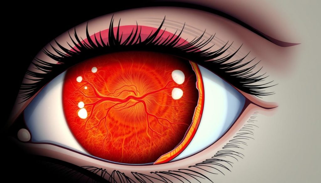
In the world of Eye Health, retinal detachment stands out as a severe concern. This guide is your first step towards understanding different Types of Retinal Detachment. It’s crucial for preventing Vision Loss. This article highlights the impact of this condition on the eye’s retina.
We aim to clarify this complicated issue and empower you with knowledge. This knowledge encourages early actions to keep your eyes healthy. Unveiling the depth of this critical matter can help prevent permanent vision loss.
What Is Retinal Detachment?
Retinal detachment is a severe condition where the retina pulls away from its underlying tissues. It’s important as the retina helps turn light into visual signals. People who are older, those with a lot of nearsightedness, or those who’ve had eye surgeries are at higher risk.

Defining Retinal Detachment in Simple Terms
Retinal Detachment happens when the light-sensitive layer inside the eye, the retina, gets separated. This stops the retina from working right and can cause Vision Problems. If not treated quickly, it might lead to lasting Eye Health issues.
The Importance of Early Detection and Treatment
Spotting a Detached Retina early is very important. Getting help fast can greatly lower the risk of serious damage and help keep your vision. If you see sudden light flashes or more floaters, you should get your eyes checked right away.
| Symptom | Potential Impact on Vision |
|---|---|
| Flashes of Light | Signal the pulling away of the retina from its position |
| Increased Floaters | Indicate bleeding inside the eye |
| Sudden Loss of Vision | Point towards severe progression of retinal detachment |
An Overview of the Human Retina and Its Function
The human retina is a key part of eye anatomy. It acts as the light-sensitive tissue at the eye’s back. Its main job is vital for vision health. It captures light and turns it into signals that the brain can understand. This process is essential for seeing.

The retina is made up of many layers with different cell types. These include photoreceptors called rods and cones. Rods help us see in low light, while cones are for color and detail in bright light. This shows how the retina adjusts to different light, showing its complexity and importance in eye anatomy.
- Light Detection: Light gets into the eye through the cornea, then the lens, and finally reaches the retina.
- Signal Conversion: The photoreceptors change light into electrical signals.
- Signal Processing: These signals are sent to the brain by the optic nerve, allowing us to see.
Problems like retinal detachment can greatly affect vision health. Knowing how the retina works shows the wonders of human biology. It also tells us how important regular eye checks are. These checks help avoid or treat problems that could harm our sight.
Types of Retinal Detachment
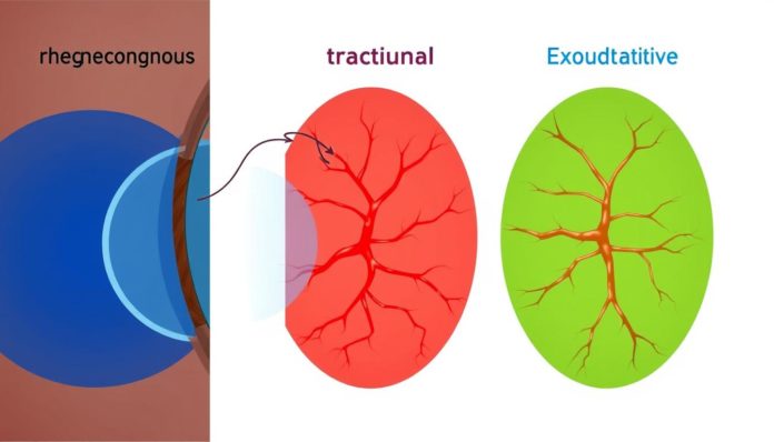
There are several forms of retinal detachment that differently impact the eye. An overview is provided using the advice from the American Society of Retina Specialists and Retina International.
Rhegmatogenous Retinal Detachment Explained
Rhegmatogenous Retinal Detachment is the most seen kind. It happens when the vitreous gel pulls away, causing tears in the retina. This lets fluid go under the retina, causing it to separate from its base.
Tractional Retinal Detachment
Tractional Retinal Detachment differs as it involves no tearing. It’s due to scar tissue on the retina that contracts. This makes the retina detach, common among diabetics.
Exudative Retinal Detachment Overview
Exudative retinal detachment happens without any tears. Fluid builds up under the retina due to inflammation or macular degeneration. These lead to leaks in the retinal layers.
| Type | Cause | Typical Outcome |
|---|---|---|
| Rhegmatogenous | Retinal tears from vitreous detachment | Retinal reattachment with timely surgery |
| Tractional | Scar tissue formation, diabetes complications | Varies; surgery might be needed to prevent vision loss |
| Exudative | Systemic diseases causing fluid leakage | Control of underlying condition stabilizes detachment |
Knowing these types helps diagnose retinal detachment correctly. It guides in making a treatment plan to avoid permanent vision loss.
Causes and Risk Factors for Retinal Detachment
Retinal detachment is a serious issue that can cause permanent vision loss if it’s not treated quickly. It’s important to know what causes it and what increases your risk. Causes include eye trauma, complications from retinal disorders, and genetic and environmental factors.
Eye Trauma: Injuries to the eye can cause retinal detachment. Accidents or sports injuries can tear the retina. When fluid gets under the retina through these tears, it can detach.
Family History: If your family has a history of retinal detachment, you might be more at risk. This shows that genetics can increase the likelihood of having retinal issues.
Age-related Eye Changes: The eye’s vitreous can shrink with age and pull away from the retina. This can create tears that lead to detachment, especially in older adults.
Being highly nearsighted, having had eye surgeries, and certain lifestyle choices can also increase your risk. Knowing these risks is crucial. If you notice sudden flashes of light or more floaters, get medical help right away.
| Cause | Description | Preventative Actions |
|---|---|---|
| Eye Trauma | Physical impact leading to tears in the retina. | Use protective eyewear during high-risk activities. |
| Family History | Genetic predisposition to retinal issues. | Regular eye examinations, especially if family history is present. |
| Retinal Disorders | Existing conditions that weaken the retina. | Manage underlying conditions with medical advice. |
| Age-related Changes | Natural degradation of the vitreous humor in the eye. | Monitor eye health regularly as age progresses. |
Understanding the causes and risks of retinal detachment is key to preventing it. It can help save your vision and lead to better outcomes for those affected by this condition.
Identifying Retinal Breaks and Tears
It’s essential to know the difference between retinal breaks and tears for eye health. Both can lead to serious issues, including decreased vision, if they’re not treated quickly and correctly.
What Leads to Retinal Breaks
Retinal breaks are often caused when the eye’s vitreous shrinks and pulls on the retina. This usually happens as we age. The vitreous may separate from the retina, leading to breaks.
Differences Between Retinal Breaks and Tears
Retinal breaks and tears are different, even though they sound similar. A break is a small hole in the retina. It might not always cause the retina to detach. A tear occurs when the vitreous pulls part of the retina, creating an opening. Fluid can then get under the retina, possibly leading to detachment. It’s crucial to treat both quickly to avoid serious issues.
| Condition | Description | Potential for Detachment |
|---|---|---|
| Retinal Break | A small hole in the retina, often without tissue loss. | Lower likelihood, but monitoring necessary |
| Retinal Tear | An actual ripping of the retinal tissue, can follow fluid accumulation. | High likelihood, immediate treatment often required |
Knowing about retinal breaks and tears can help prevent retinal detachment. Regular eye exams are key, especially if you have symptoms like vitreous detachment. Early detection is vital. This can prevent the need for complex treatments like retinal surgery.
Symptoms Indicating a Possible Retinal Detachment
Noticing the early signs of retinal detachment is key to avoid losing your vision. We will look at important visual symptoms you should not ignore. These signs are crucial to watch out for.
Visual Signs You Shouldn’t Ignore
If your vision suddenly gets worse or you see a lot more floaters, get medical help right away. Blurry vision is also a major alarm that may suggest your retina is detaching. These warnings might start off small but can quickly worsen, leading to permanent damage if not treated fast.
Understanding Flashes and Floaters
Many people see flashes of light or more floaters, indicating their retina might be detaching. These signs can look like brief lightning flashes or tiny black or grey dots moving in your sight. While having a few floaters is normal, a sudden increase with flashes means you should act fast to check for retinal detachment.
- Sudden Vision Loss: Instant decrease in vision, appearing as if a curtain has fallen across your eyesight.
- Visual Floaters: A rise in tiny shapes floating in your view, looking like thin threads or small specks.
- Light Flashes: Unexpected flashes of light, mostly seen on the sides of your vision, not caused by outside lights.
- Blurry Vision: Blurred vision that starts suddenly or slowly, making it hard to do everyday tasks.
Knowing these symptoms and their importance for your retina’s health is vital. If you see any of these signs, especially more than one, you should contact a healthcare provider right away.
Diagnostic Procedures for Retinal Detachment
When diagnosing retinal detachment, various highly effective methods are key. Each one helps doctors accurately find and measure the detachment. This guides them in choosing the right treatment. Below are the main diagnostic tools used.
Eye Examination: This first step is a detailed eye exam. Ophthalmologists look inside the eye carefully. They check for any signs that could mean retinal detachment.
Retinal Imaging is crucial for getting clear pictures of the retina. Tools like fundus photography and optical coherence tomography (OCT) are used. They make it easier to spot any detached parts or tears in the retina.
Ophthalmoscopy offers a live view of the retina. With an ophthalmoscope, the doctor checks the back of the eye closely. They can see any tears or places where the retina has detached.
Ultrasound comes in handy when ophthalmoscopy can’t give clear enough pictures, for example, if the patient’s lens is cloudy. Using ultrasound waves, doctors can see pictures of the eye’s inside. This shows them any parts of the retina that are detached.
These diagnostic tools are not just important for finding retinal detachment. They’re also vital in figuring out how serious it is and exactly where it’s located. This directly helps choose the best treatment.
| Diagnostic Tool | Function | Use Case |
|---|---|---|
| Eye Examination | Initial inspection and detection of physical and structural abnormalities in the retina. | Basic screening to identify unusual signs and symptoms related to retinal health. |
| Retinal Imaging | Acquires detailed visual representations of retinal layers. | Used to identify the specific type and extent of retinal detachment. |
| Ophthalmoscopy | Enables a direct view of the retina, allowing diagnosis of tears and detachment locations. | Critical for visual assessment during follow-up evaluations. |
| Ultrasound | Generates imaging when direct visualization is obstructed. | Applied in cases where optical clarity is compromised by conditions like cataracts. |
With these diagnostic methods, medical experts can quickly and accurately detect retinal detachment. This sets the foundation for the right treatment.
Emerging from the Shadows: Posterior Vitreous Detachment
Posterior Vitreous Detachment (PVD) is common but often not well understood. It’s very important for our Retinal Health. Knowing about this helps to catch early signs of Proliferative Vitreoretinopathy and other eye problems.
The Relationship Between PVD and Retinal Detachment
PVD happens when the vitreous, a gel inside the eye, pulls away from the retina. This can sometimes be harmless. But it can also lead to dangerous retinal tears. If these aren’t treated, the retina can fully detach. It’s key to catch PVD early to stop Proliferative Vitreoretinopathy. This condition can greatly harm your vision.
Recognizing Posterior Vitreous Detachment Symptoms
Seeing flashes of light, floaters, and shadows are symptoms of PVD. If you notice these, talk to an eye doctor right away. This can help protect your Retinal Health.
| Condition | Common Symptoms | Potential Complications |
|---|---|---|
| Posterior Vitreous Detachment | Flashes, floaters, visual shadow | Retinal tear, Detachment |
| Proliferative Vitreoretinopathy | Visual distortion, decreased vision | Severe retinal detachment |
Proliferative Vitreoretinopathy: Complication of Retinal Detachment
Proliferative Vitreoretinopathy (PVR) often follows retinal detachment. It poses a big challenge to Vision Restoration. PVR leads to Scar Tissue growth inside the eye. This can pressure the retina, causing more visual problems and making recovery harder.
When we talk about Retinal Surgery Complications, PVR stands out. It complicates surgery and affects the chance of getting one’s vision back. The scar tissue can tighten and may cause the retina to detach again. This might need more surgeries, from small fixes to bigger operations.
Understanding how PVR works and what causes scar tissue is key. It helps find better treatments and improve surgery results for people getting their retina attached again.
- Spotting the condition early and acting fast to control scar tissue growth.
- Better surgical methods that reduce harm and movement to tissues.
- Care after surgery that focuses on reducing inflammation, which leads to Scar Tissue.
Stopping PVR early is key to better Vision Restoration rates. It also lowers the risk of the retina detaching again. That’s why improving drugs and surgery tools is crucial in fighting against PVR and other major Retinal Surgery Complications.
| Complication | Impact on Surgery | Importance in Recovery |
|---|---|---|
| Proliferative Vitreoretinopathy | Can necessitate repeated surgeries | Crucial for Visual outcomes |
| Scar Tissue Formation | Challenges in achieving successful attachment | Critical for long-term stability of retina |
| Inflammation | Must be tightly controlled | Essential for healing and preventing further PVR |
To fight Proliferative Vitreoretinopathy, we need a mix of good surgery plans, ongoing research, and careful after-surgery care. This will help us improve how we fix retinas and help people see again.
Treatment Options for Retinal Detachment
Retinal detachment is a serious condition that threatens sight. Luckily, medical advances have brought about effective treatments. Institutions like the American Academy of Ophthalmology and the Cleveland Clinic recommend key surgeries. These include Laser Photocoagulation, Cryotherapy, Scleral Buckling, and Vitrectomy.
Laser Surgery and Cryopexy
Laser Photocoagulation and Cryotherapy are often first in line for fixing retinal breaks. Laser Photocoagulation uses a precise laser beam to create scarring. This scarring helps weld the retina to the underlying tissue. Cryotherapy uses extreme cold to form a similar scar. This scar attaches the retina to the eye wall.
Scleral Buckling Procedure
Scleral Buckling is great for torn or broken retinas. It involves placing a flexible band around the eye’s white part (sclera). This acts against the force pulling the retina out of place. Even though it sounds complex, Scleral Buckling is highly effective. It has been a key method for fixing certain tears and detachments for years.
Vitrectomy: An In-Depth Look
In severe cases, doctors may opt for a Vitrectomy. This involves removing the vitreous gel. The aim is to stop it from pulling on the retina. The space is then filled with saline, gas, or silicone oil. This helps the retina stay in place. A Vitrectomy is considered when there’s a lot of scarring. It’s also an option if surgeries like Laser Photocoagulation or Cryotherapy didn’t work.
This table outlines when to use each Retinal Surgery:
| Treatment | Indicated for | Considerations |
|---|---|---|
| Laser Photocoagulation | Small tears without detachment | Minimal recovery time, outpatient |
| Cryotherapy | Localized detachment, accessible tears | Could induce inflammation |
| Scleral Buckling | Larger or multiple tears | More invasive, longer recovery |
| Vitrectomy | Complex, advanced PVR cases | Potential additional surgeries |
Life After Retinal Surgery
Retinal surgery is a key step in keeping your vision healthy. What you do after surgery is just as important. Knowing what to expect helps patients through this time.
Recovery Expectations and Guidelines
After retinal surgery, taking care of yourself is vital. Patients might feel a bit uncomfortable or see things oddly at first. It’s very important to follow your doctor’s advice closely to avoid any problems.
Be sure not to do heavy activities, keep your eyes dry, and use eye drops as told. These steps help stop infections and reduce swelling.
Long-Term Care and Monitoring
Checking your retinal health after surgery is key to know if the surgery worked and to keep your retina healthy. Going for regular visits helps spot issues like retinal re-detachment or new cataracts quickly. Sometimes, therapy to improve your sight and help you get used to any vision changes might be needed.
| Post-Surgery Timeline | Important Actions | Expected Outcomes |
|---|---|---|
| 1-2 Weeks | Avoid heavy lifting and swimming | Prevention of physical stress on the eyes |
| 1 Month | Follow-up visit for eye examination | Assessment of surgical success and healing |
| 3-6 Months | Regular vision tests and retina scans | Ongoing monitoring for stable vision and retinal health |
Proper care after surgery and going for check-ups help a lot in recovering and keeping your eyes healthy. Making sure you get medical care when needed and paying attention to your vision is essential for your ongoing wellness.
Preventing Retinal Detachment Through Lifestyle Adjustments
Living a healthy lifestyle can greatly lower your risk of retinal detachment. It is crucial to focus on eye protection, regular eye exams, and nutritional supplements. To boost eye health, you need to take steps before problems start.
Making certain lifestyle and diet changes can help strengthen your eyes. We’ll share tips and nutrients that top health groups recommend.
According to Harvard Health Publishing, regular physical activity and eating well can prevent eye problems that lead to retinal detachment.
- It’s vital to wear sunglasses that block UV rays for eye protection.
- Getting regular eye exams helps find early retina issues, avoiding serious damage.
- Nutritional supplements with vitamin C, E, and zinc can maintain retinal and eye health.
- Eating foods like leafy greens and omega-3s is good for your eyes’ health and structure.
Mixing these actions can offer a strong guard against retinal detachment.
Following a healthy lifestyle benefits your eyes and overall health. It greatly helps keep your vision and body in good shape.
| Lifestyle Factor | Impact on Eye Health |
|---|---|
| UV-Protective Eyewear | Shields retina from UV damage |
| Regular Eye Check-ups | Early diagnosis of potential retinal issues |
| Diet rich in Vitamins and Minerals | Supports retinal durability and prevents degeneration |
Integrating healthy lifestyle habits is key to protecting your vision. While everyone’s needs differ, combining protection, diet, and medical care can prevent serious eye issues like retinal detachment.
Support and Resources for Individuals with Retinal Detachment
Retinal detachment can be tough, both on your body and mind. Luckily, there’s a lot of support out there. Getting to know eye health resources, joining patient support groups, and getting involved in the retinal detachment community are key. These steps help manage and beat the challenges of this condition.
Patient Support Groups: These groups are a place to share stories, fears, and experiences. They offer comfort and advice from people who really get what you’re going through. Being part of these groups reduces loneliness and worry. It builds a supporting network that brings hope and strength.
Eye Health Resources: Many organizations offer current, research-based details on retinal detachment. This information helps educate patients and their families about the condition, treatments, and recovery expectations. Knowledge gives power. Access to accurate information empowers patients and their caregivers greatly.
Retinal Detachment Community: Joining the wider retinal detachment community opens doors to medical insights and daily living tips with vision loss. Community events, webinars, and forums are organized for members to connect. They share resources and support each other emotionally and physically.
- Ask healthcare providers about patient support groups and eye health resources.
- Join online forums and social media groups focused on retinal detachment. These help you keep up with treatments and share experiences.
- Look for public health websites and eye care institutions for informative materials.
- Go to health fairs and eye health seminars. There, you can meet professionals and others with similar issues.
Being active in these support groups helps you and others facing similar problems. Recovery and daily life management is a journey. The right support makes this journey easier.
The Future of Retinal Detachment Treatment
The field of Retinal Research is filled with exciting updates. These updates promise big changes in how we treat retinal detachment. Journals like ‘Journal of Clinical & Experimental Ophthalmology’ and ‘Future Ophthalmology’ share updates about Innovative Eye Treatments. These treatments will likely make patient recovery better than ever. Thanks to Medical Advancements, future eye care will bring hope and better vision restoration.
Scientists are working on new ways to fix the retina with great accuracy. They use advanced imaging and new materials. They are also looking into regenerative medicine, like stem cell therapy. This could not only fix but regenerate damaged retinal cells. We’re moving towards a future where we might fully repair the retina.
Looking forward, there’s a stronger emphasis on care that focuses on the patient. This means treatments that are easier and less invasive. Experts are aiming for discoveries that allow quick recoveries and less risk. The progress in eye care shows the dedication to improve the lives of those with retinal issues.
FAQ
What are the different types of retinal detachment?
Retinal detachment comes in three types: rhegmatogenous, tractional, and exudative. Each type has its own causes and ways to treat it.
How can I tell if I might have a retinal detachment?
Signs you might have a retinal detachment include sudden light flashes and new floaters. You might also see a shadow over your vision or lose vision suddenly. If you see these signs, get medical help right away.
What is the function of the human retina?
The retina works by sensing light at the eye’s back and sending pictures to the brain. This process helps you see.
How urgent is it to treat a detached retina?
Treating a detached retina is very urgent. Not treating it can cause you to lose your vision forever. It’s important to find and treat it early on.
What leads to retinal breaks, and how are they different from retinal tears?
Retinal breaks happen when the retina gets thin and weak. Tears happen when the vitreous pulls on the retina hard. Both can lead to detachment if not treated.
Can retinal detachment be prevented?
You can’t prevent all causes of retinal detachment, like aging or family history. But, protecting your eyes and living healthily can lower your risk.
What does recovery from retinal surgery involve?
Recovery time from retinal surgery varies but usually takes weeks. It’s key to follow your doctor’s advice and go to all check-ups after surgery.
What is the relationship between posterior vitreous detachment (PVD) and retinal detachment?
PVD occurs when the eye’s vitreous gel separates from the retina. This can lead to retinal tears and detachment, especially if the vitreous sticks too much to the retina.
What are the treatment options for retinal detachment?
Treatments include laser surgery and cryopexy to fix tears or holes, scleral buckling to support the retina, and vitrectomy to remove vitreous gel and fix the retina.
Are there any new treatments for retinal detachment on the horizon?
Yes, research into better surgical techniques and imaging improvements is ongoing. This could introduce new ways to treat retinal detachment soon.
What lifestyle adjustments can help prevent retinal detachment?
To help prevent detachment, keep a healthy lifestyle, protect your eyes, get regular check-ups, and consider nutritional supplements to support eye health.
Where can I find support if I’ve been diagnosed with retinal detachment?
Support can be found through patient groups, eye health organizations, and online communities. They provide resources for those dealing with retinal detachment and vision issues.


