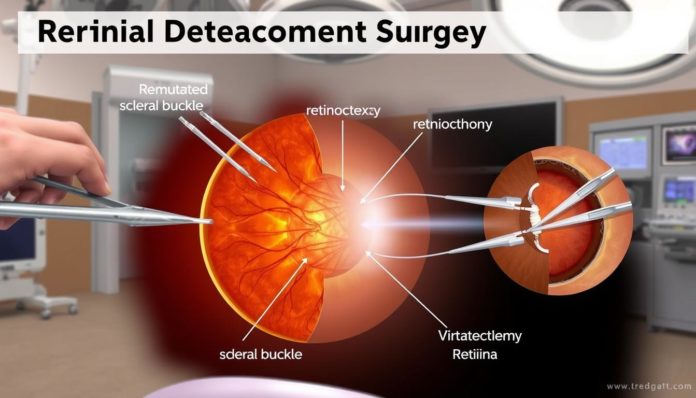
Getting told you have a detached retina is worrisome. It sparks a lot of questions about how to keep your eyes healthy. Knowing about detached retina surgery options is key in making wise choices. This article will guide you through the different ways doctors can fix your vision. We’ll explore how each method works to get your eye health back on track.
Introduction to Retinal Detachment
What is retinal detachment? It’s when the retina separates from the back of the eye. This can lead to vision loss if not treated right away. Knowing about this condition is key because it shows how much you need quick retinal detachment treatment.

Spotting and treating this issue early can stop vision damage. Sudden symptoms like increased floaters, light flashes, or a shadow in your vision are warnings. If you notice these, get medical advice right away to talk about retinal detachment treatment options.
Here’s why immediate action is important:
- Prevention of permanent vision loss: Waiting too long for treatment could mean losing your sight for good. Quick action is key.
- Increased treatment options: Finding the problem early means you might get less invasive treatments. This could lead to better recovery results.
- Reduction of healthcare costs: Quick treatment can prevent the need for more complex surgeries.
We want to highlight how vital it is to know the signs of retinal detachment. Understanding what is retinal detachment helps you recognize symptoms early. Then, you can get the help you need promptly.
What Causes a Retina to Detach?
It’s key to know why retinal detachment happens to stop it early. Many things cause this serious eye issue, which might lead to lost sight if uncured. Being older, hurt, diabetic, or very nearsighted ups your risk.

When hit by something or doing intense sports, the retina might quickly separate from its base. This messes up the eye’s vital layers. As we age, the eye’s gel might shrink and pull away. This can tear the retina and lead to detachment.
If you’re really nearsighted, your eye stretches and thins the retina, making it weak. Diabetes makes things worse by changing blood vessels. This change can cause diabetic retinopathy, moving on to retinal detachment.
| Condition | Impact on Retina | Prevalence |
|---|---|---|
| Trauma | Immediate separation through force | Variable, associated with severe impacts |
| Aging | Vitreous shrinkage causing tear | Highly common in elderly adults |
| High Myopia | Stretching and thinning of retina | Common in severly nearsighted individuals |
| Diabetes | Blood vessel alterations leading to retinopathy | Common in long-term diabetics |
To prevent retinal detachment, get your eyes checked often, especially if you’re at risk. Handle any health problems that could make your eye health worse. Keeping an eye out and managing these issues helps lower your chances of retinal detachment.
Signs and Symptoms of Retinal Detachment
Knowing the signs of retinal detachment symptoms early can make a big difference. If ignored, it can cause major vision loss or even blindness. So, spotting a detached retina soon is key.
- Blurred vision that gets worse over time
- Flashes of light in one or both eyes
- More floaters—small, dark spots or lines floating in your vision
- A shadow or curtain over part of your vision, which seems to come down from the top or moves in from the side
Seeing an eye expert right away is crucial if you notice any of these signs. Acting fast can mean saving your sight.
| Symptom | Description | Urgency |
|---|---|---|
| Flashes of Light | Unexpected flashes like camera flashes | High |
| Floaters | Tiny, moving dark shapes in your view | Moderate to High |
| Vision Shadowing | A dark cover or shadow blocking your view | Immediate |
This table shows crucial retinal detachment symptoms and the need for quick action on detecting a detached retina. Treat every listed symptom as a possible emergency that needs fast attention.
Evaluating the Severity of Retinal Detachment
Understanding how serious a retinal detachment is means getting an accurate eye check. Eye experts use different tools to look at the eye closely. This helps not only in finding out what’s wrong but also in making a good plan to treat it.
Assessing retinal detachment is about closely looking at the eye with special gear. They aim to see how much of the retina is affected and where. Knowing this decides how urgent the treatment is and what kind it should be.
- Ophthalmoscopy: This equipment lets the doctor peek through the pupil to the retina. This way, they can tell how apart the retina is.
- Ultrasound imaging: If ophthalmoscopy doesn’t give clear results, ultrasound imaging can offer more info about the eye’s back.
- Optical coherence tomography (OCT): This safe imaging test gives clear pictures of the retina. It shows which layers are detached.
Finding out more about retinal detachment through these methods is key. It helps decide on quick and right action. Being quick and correct with the eye exam can really help the healing process.
It’s very important to know how each tool and method works during the eye exam. Every step helps understand better, which is vital for safekeeping the patient’s sight. This ensures they get the best care possible.
Retinal Detachment Surgery: An Overview
Retinal detachment needs various surgeries for fixing vision and eye health. We will look at the main procedures used to treat this serious issue.
Scleral Buckle Surgery Explained
Scleral buckle surgery puts a silicone band around the eye. This helps hold the retina in place. It’s mainly used when there are many tears or a big detachment, making the retina stay against the eye wall.
Vitrectomy for Retinal Detachment: The Procedure
In a vitrectomy, doctors remove the vitreous gel that can pull on the retina. Then, they fill the space with saline, silicone oil, or gas to keep the retina fixed while it heals. This is great for serious or complicated detachments.
Pneumatic Retinopexy: How It Works
Pneumatic retinopexy is simpler and done outside a hospital. It injects a gas bubble into the eye. The patient must keep their head in a certain angle so the bubble helps reattach the retina. This is combined with cryopexy or laser to ensure the retina stays in place.
Comparing Retinal Detachment Treatment Options
Choosing the right retinal detachment repair method is key for good recovery and vision results. We’ll look at common treatment options, comparing their invasiveness, recovery times, and success rates. These factors are vital for treatment comparability.
Each treatment option for retinal detachment brings its own benefits and things to consider. The table below shows a comparison of the three most used surgeries in retinal detachment repair.
| Treatment Method | Invasiveness | Recovery Time | Success Rate |
|---|---|---|---|
| Scleral Buckling | Moderate | 2-4 weeks | Approx. 85% |
| Vitrectomy | High | 4-6 weeks | Approx. 90% |
| Pneumatic Retinopexy | Low | 1-2 weeks | Approx. 70% |
The data above is essential for making informed choices. It shows the major differences between treatment options. Knowing this helps patients and doctors decide based on the detachment’s severity and the patient’s health. Understanding these procedures makes choosing the right retinal detachment repair easier.
Customizing Retinal Detachment Repair to Patient Needs
The changes in individualized retinal treatment and personalized eye surgery have greatly improved how doctors treat unique patient needs in retinal detachment. Now, understanding that everyone’s eye health is different, surgeons focus on custom solutions. These solutions consider the patient’s medical history, the seriousness of their condition, and how it affects their life.
Surgeons now use advanced tools to create personalized eye surgery plans. These plans not only aim to fix the retinal detachment but also help patients recover better. The patient’s age, how long the retina has been detached, and other eye problems are important in choosing the right surgery.
| Patient Factor | Impact on Surgical Approach | Suggested Surgical Options |
|---|---|---|
| Age | Younger patients might recover faster and can handle more invasive procedures. | Vitrectomy, Scleral Buckling |
| Duration of Detachment | Longer durations may require more complex surgeries to ensure complete attachment and healing. | Vitrectomy with Laser Photocoagulation |
| Additional Eye Conditions | Presence of conditions like diabetic retinopathy may modify the type and extent of surgery needed. | Pneumatic Retinopexy, Modified Vitrectomy |
The aim of individualized retinal treatment is not just fixing the retina. It’s about improving the patient’s life with as little pain and as much efficiency as possible. Medical technology and surgery methods are always getting better. This means care in retina surgeries is more personalized than ever, focusing on what each patient needs.
Understanding the Risks of Retinal Detachment Surgery
Retinal detachment surgery helps fix vision but has risks. We’ll look at the risks of retina surgery, common retinal surgery complications, and what to expect for vision surgery prognosis.
Potential Complications from Surgery
Although usually successful, retinal detachment surgery can have complications. Some issues include:
- Infection that might require more treatment.
- Bleeding within the eye, which can worsen vision.
- Retinal re-detachment, meaning the retina detaches again.
- Cataract formation, often needing more surgery.
These retinal surgery complications may lead to more surgeries. They can also affect recovery speed and comfort.
Long-Term Outlook After Surgery
The outcome after retinal detachment surgery depends on several things. It matters how bad the detachment was, how quickly you got treatment, and your health. Many patients see a big vision improvement but full recovery could take months.
Visual acuity might not completely return to what it was. This is especially true if the retina was detached for a long time before surgery. It’s important to have regular check-ups to watch healing and handle any issues.
Knowing these risks and keeping realistic expectations can help. Being well-informed before surgery and having a good recovery plan are key for the best outcome.
Preparing for Detached Retina Surgery: Steps to Take
Getting ready for retina surgery means you need to take some important steps. These steps help you prepare for both the surgery and the recovery after. Having a good pre-surgery checklist helps lower risks and makes the surgery more successful.
Let’s look at the main things every patient should do.
- Medical Evaluation: Get a full medical check-up to understand the retinal detachment and any issues that could impact the surgery.
- Diet and Medication: Change your diet as your doctor suggests. Talk about any current medicines to see if you should stop them for a while.
- Arranging Transportation and Aftercare: You won’t be able to drive right after the surgery. So, make plans for someone to drive you home. Also, set up help for the days following the surgery.
- Pre-operative Fasting: Follow your surgeon’s fasting rules to get ready for the anesthesia.
Talking to your healthcare provider is key. It helps you get rid of doubts about the surgery. It’s also good for understanding every step of the surgery and the healing after.
| Checklist Item | Description | Preparation Needed |
|---|---|---|
| Consultation | Talk about your general health and your retinal issue | Make a time to see a retina expert |
| Dietary Adjustments | Change your diet for the best health before surgery | If needed, see a nutritionist |
| Medication Review | Go over all your current medicines with the doctor | Give a full list of your meds to the surgeon |
| Transport & Care | Set up rides and help for after the surgery | Get a friend or family member ready to assist |
Following all these steps is key when getting ready for retina surgery. This pre-surgery checklist organizes how you get ready. It makes the surgery and healing smoother and more successful.
Retinal Detachment Surgery: The Recovery Process
Recovery after retina surgery means following certain rules closely. It’s important to watch for signs that show healing is going well. Knowing what to do after surgery helps patients get better results.
Post-Surgery Care Guidelines
The time just after retina surgery is very important. Patients should not do anything too hard that could harm the healing eye. This means not lifting heavy things or playing tough sports.
Taking care of the eye, keeping it clean, and taking all medicines as told are key for healing.
Monitoring for Signs of Successful Healing
Good signs of healing include less distortion in vision or not seeing new floaters. Making sure to go to all doctor’s visits is crucial. These check-ups help see how well the retina is healing and catch any problems early.
By following these steps, patients can help their recovery and get back to normal activities with better vision.
Advancements in Laser Surgery for Retinal Detachment
The latest in retina laser surgery has greatly improved eye care, especially for retinal detachment. Thanks to technological progress in eye surgery, patients now benefit from less invasive and more efficient procedures.
Micro-pulse lasers are at the forefront of these innovations. They repair the retina without harming nearby tissue. This approach reduces recovery time and improves the results of surgery.
Another breakthrough is the use of real-time imaging systems in surgery. These systems help surgeons operate with incredible precision. This reduces risks and makes complex procedures routine. The technological progress in eye surgery is making surgery safer and more accurate.
| Technology | Benefits | Impact on Recovery Time |
|---|---|---|
| Micro-Pulse Laser | Minimizes tissue damage | Reduces recovery period significantly |
| Real-Time Imaging Systems | Increases surgical accuracy | Decreases risk of additional surgeries |
The latest in retina laser surgery doesn’t just offer better vision outcomes. It also helps us learn more about retinal health. The technological progress in eye surgery is transforming difficult surgeries into simpler outpatient procedures.
Retinal Detachment Surgery Success Rates
Exploring the success rates of retina surgery is very important. It offers valuable insights for both patients and doctors. We’ll look into what affects these rates and show key statistics.
Factors Influencing Surgical Outcomes
The success of retina surgery can really vary. It depends on how severe the detachment is, the surgery method, the surgeon’s skill, and the patient’s health. Knowing these factors can help improve success rates of retina surgery.
Statistical Data on Retinal Repair Success
To give a clear view of outcomes of retinal repair, here’s data on success rates. It shows how different methods and conditions have fared:
| Technique | Success Rate | Timeframe |
|---|---|---|
| Scleral Buckle | 90-95% | 1 Year |
| Vitrectomy | 85-90% | 1 Year |
| Pneumatic Retinopexy | 70-75% | 1 Year |
This table shows the impact of various surgical techniques on success rates of retina surgery. It’s vital for making the right choice about retinal repair methods.
Insurance Coverage and Cost Considerations for Retinal Surgery
Grasping the costs of retinal detachment surgery is key for those getting ready for it. Costs can be influenced by insurance for eye surgery and personal expenses. This can greatly affect the total cost.
The retinal detachment surgery cost differs based on the surgery type and how severe the retinal detachment is. Patients should talk to their doctors about the best treatments and their costs.
Most health insurance plans help pay for some retinal surgery types, but coverage varies. It’s crucial for patients to check their insurance benefits. They’ll learn what cost they must cover, including deductibles, copays, and coinsurance.
- Contact your insurance provider to confirm coverage details for retinal surgeries.
- Ask your healthcare provider about possible payment plans or financial assistance for out-of-pocket expenses.
- Consider additional costs for post-surgery medications and follow-up visits, which may not be covered by insurance.
Having insurance for eye surgery can take away much of the financial stress. Yet, patients must actively manage their expenses and know their coverage limits. The aim is to avoid surprises and make sure money issues don’t block needed healthcare.
When to Seek a Second Opinion for Retinal Detachment Procedure
Decisions about eye surgery should not be made in haste, especially with retinal detachment. It’s crucial that the procedure fits your specific needs. Seeking a second opinion on retina surgery can be helpful.
Getting another view can offer new insights or different treatment options better suited for you. It’s wise if you’re unsure about your first diagnosis or treatment plan.
Seeing another eye surgeon reflects your carefulness about your vision, not mistrust in your doctor. With complex retinal detachment cases, multiple surgical options might exist. Understanding each one’s benefits and risks is key.
An extra opinion can introduce you to new techniques and possibly suggest a less invasive method. This helps you make a more informed choice.
Every person’s eye condition is unique. Your overall health, lifestyle, and vision needs affect which surgery is best. Consulting another surgeon lets you customize the surgical approach.
This step can confirm the first recommendation or open up new possibilities. Before such a significant operation, a second opinion provides the confidence you need.
FAQ
What are the different types of retinal detachment surgery available?
A: Scleral buckle surgery, vitrectomy, pneumatic retinopexy, and advanced laser surgery are the main types. Each one is chosen based on the retinal detachment’s severity and type.
How do I know if I have a retinal detachment?
Seeing sudden floaters, flashes of light, or a shadow in your vision are common signs. If you notice these, see a doctor right away. Early action is critical.
What causes the retina to detach?
A retinal tear or hole, eye injury, severe diabetes, or inflammatory issues can cause detachment. As we get older, changes in the eye might also lead to it.
Are there any risks associated with retinal detachment surgery?
Retinal surgery might lead to infection, bleeding, or increased eye pressure. There’s also a chance of cataract formation. Sometimes, vision might not fully return. Your doctor will talk about these risks before surgery.
How do doctors determine the severity of a retinal detachment?
Doctors use a detailed eye exam to figure out how bad the detachment is. They might use tools like ultrasound or OCT. They look at the detachment’s location, size, and more to plan treatment.
What is scleral buckle surgery?
Scleral buckle surgery involves putting a silicone band around the eye. It presses the eye wall against the retina. This helps close breaks and fix the retina.
What does vitrectomy for retinal detachment entail?
In a vitrectomy, the eye’s vitreous gel is taken out. The retina is then fixed using laser surgery or cryotherapy. The eye is filled with gas or silicone oil to keep the retina in place as it heals.
How does pneumatic retinopexy work?
Pneumatic retinopexy uses a gas bubble injected into the eye. The bubble helps press the retina back in place. Then, laser or freezing treatment seals the tear.
What surgeries are available for treating retinal detachment?
Treatments include scleral buckle surgery, vitrectomy, and pneumatic retinopexy. Often, these are combined with laser surgery to fix tears or holes.
Can personalized factors affect the choice of retinal detachment surgery?
Yes. The surgery choice depends on how bad the detachment is, the health of the eye, age, and lifestyle. An eye surgeon will think about these when planning the repair.
How do I prepare for detached retina surgery?
You might need to stop some medicines, arrange a ride home, and fast before the surgery. Follow your doctor’s pre-surgery instructions closely.
What is involved in the recovery process after retinal detachment surgery?
After surgery, rest your eyes and avoid hard activities. Use any prescribed eye drops and watch for healing signs. You’ll also have check-ups to see how you’re healing.
What advancements have been made in laser surgery for retinal detachment?
Recent tech has made laser surgery more precise and less invasive. This leads to faster recovery and better success rates.
What are the success rates for retinal detachment surgery?
The success rates are generally high but vary by case. Quick treatment, overall eye health, and the surgeon’s skill matter.
How is retinal detachment surgery covered by insurance, and what are the cost considerations?
Insurance usually covers necessary surgery, but coverage differs. Understand your policy, your costs, and any extra charges for follow-up treatments.
When should I consider getting a second opinion for a retinal detachment procedure?
If you’re not sure about the treatment plan, if it’s a complex case, or you just want more info, a second opinion can be helpful. It might offer more options.


