When the inner linings of our eyes come loose, our vision isn’t the only thing at risk. Suddenly, an entire world might fade into darkness. This happens with retinal detachment, a serious issue that needs quick, expert treatment in ophthalmology. It happens when the retina loses its connection to the tissue supporting it, needing medical help to stop lasting vision loss. Scleral buckling steps in as a trusted solution in this battle—a beacon of hope for those on the brink of major sight loss.
Experts in eye health widely recommend scleral buckling. It’s more than just an eye surgery; it’s a mission to restore visual harmony. Picture it as a gentle hug: a custom-fit silicone band wraps around the eye. It carefully pushes the sclera to reconnect with the retina. This process is a fine dance of skill and know-how, aiming to resurrect our ability to see the world clearly.
Looking closer at this impactful operation, remember that scleral buckling is more than a method. It’s a lifeline for those facing retinal detachment. We’re taking a trip into the world of ophthalmology, exploring the wonders of modern eye surgery. For anyone dealing with such a condition, learning about this surgery is the first sign of hope. It’s a light starting to shine in what seemed like a permanently dim world.
What is Retinal Detachment and Why It’s Serious
Retinal detachment is seen by experts, like those at the American Academy of Ophthalmology, as very serious. It happens when the retina moves from its place at the back of the eye. This leads to subretinal fluid building up. Without treatment, it can cause a big loss in how well you see.
Symptoms can start suddenly. You might see flashes of light, more floaters, and a shadow across your vision. Knowing these signs and catching the problem early is very important. It helps stop worse damage to your sight.

It’s vital to understand how serious retinal detachment is. When the retina detaches, it can’t send visual info from the eye to the brain right. This messes up how you see, leading to symptoms and possibly long-term damage.
| Symptom | Potential Consequence | Urgency Level |
|---|---|---|
| Flashes of Light | Early sign of retinal tearing | High |
| Increased Floaters | Possible retinal detachment | Immediate |
| Shadow over vision | Advanced retinal detachment | Critical |
If you notice any of these symptoms, seeing a doctor right away can really help. It can lead to better results and protect your visual acuity. Quick understanding and action are crucial. They ensure you get the right treatment fast.
Introduction to Scleral Buckling
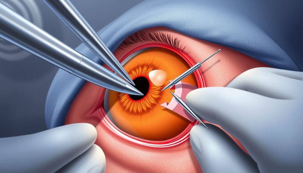
Scleral buckling is key in eye surgery, tackling retinal detachments. It uses a silicone band placed around the eye. This band gently pushes the eye’s wall against the retina. The aim is retinal reattachment, to fix the eye’s shape and function.

This surgery mainly uses a silicone band. It wraps around the eye’s white part (sclera). It helps lessen retina stress, aiding in fixing the retina. This is crucial to restore clear vision.
How Scleral Buckling Works
The scleral buckling method involves attaching a silicone band surgically. This band indents the eye, easing tension on the retina. It encourages the retina to stick back to its base. Cryotherapy may also be used to close any tears, helping recovery.
The Role of Scleral Buckling in Eye Surgery
In eye surgery, scleral buckling is prized for fixing retinal detachments less harshly. It not only aids in retinal reattachment but also helps save, even improve, vision. Because it’s effective and safe, it’s a key method for treating retinal detachments.
Determining the Need for Scleral Buckling
Figuring out if scleral buckling is needed for retinal detachment starts with a full eye examination. A skilled vitreoretinal specialist does this. It’s vital for precisely diagnosing and managing eye problems.
Various tools help gauge the retinal detachment during the exam. These include high-tech imaging for a clear inside look at the eye.
- Dilated eye exams give the specialist a wide view of the retina.
- Ultrasound imaging helps show how far the retina has detached.
- Optical Coherence Tomography (OCT) captures detailed retina images, identifying areas needing repair.
This detailed check-up helps the vitreoretinal specialist decide on the right action. They look at the retinal detachment’s specifics, considering the patient’s eye health.
The final choice to use scleral buckling is based on the case details. The specialist aims for the best treatment outcome, focusing on restoring vision effectively.
Types of Retinal Detachments Treated with Scleral Buckling
Scleral buckling is a key surgical approach in ophthalmology. It’s highly effective for various retinal detachment types. The procedure is vital for handling both rhegmatogenous and tractional retinal detachment. These are treated with different techniques to meet each case’s specific needs.
Rhegmatogenous retinal detachment is pretty common. It happens when the retina breaks or tears. Fluid then builds up underneath. The retina starts to peel off from its support tissue. The scleral buckling technique stops these breaks. It prevents the retina from detaching further by pushing the sclera inward.
Tractional retinal detachment is less common. It’s often linked to diabetes, which causes scar tissue to form on the retina. This scar tissue then contracts, making the retina pull away. Scleral buckling can help. It eases the traction on the retina. But, doctors must be extra careful with the delicate retinal tissue.
| Detachment Type | Common Causes | Treatment Approach |
|---|---|---|
| Rhegmatogenous | Breaks or tears in the retina | Scleral buckling to seal retinal breaks and support tissue adhesion |
| Tractional | Diabetic retinopathy, scar tissue formation | Selective use of scleral buckling to reduce traction |
Knowing the difference between retinal detachment types shows the need for scleral buckling. Each type needs a careful checkup to plan the right surgery. This is why scleral buckling works well in fixing the retina.
Pre-Surgical Considerations for Scleral Buckling
Getting ready for scleral buckling means taking some key steps. These steps can really help your eye surgery go well. Here’s what you should remember:
- It’s vital to talk with an eye specialist about your full medical history.
- You may need to stop taking some meds that could mess up the surgery. These might cause extra bleeding or interact with anesthesia.
- You’ll likely have some lab tests to check if you’re in good shape for surgery.
- An in-depth eye exam is essential to look at the retina and other parts of the eye.
- Make sure to tell your doctor about any allergies, especially to meds or stuff used in surgery prep.
Here’s a quick guide on getting ready for scleral buckling:
| Preparation Aspect | Description | Importance |
|---|---|---|
| Medical Consultation | Talk about your health history and any meds you’re taking. | Makes sure the surgery plan is safe and right for you. |
| Laboratory Tests | May include blood tests among other necessary tests. | Checks if you’re healthy enough for the procedure. |
| Eye Examination | A thorough check-up by an eye doctor. | Important for deciding on the best approach for surgery. |
Scleral buckling is a delicate procedure that needs careful eye handling. If you prepare properly, you up your chances for a good result and easier healing.
The Scleral Buckling Procedure Explained
The scleral buckling procedure is a key technique in eye surgery for fixing retinal detachment. It brings hope and vision restoration to patients. The procedure is delicate, needing careful work and top-notch materials for success.
Step-by-Step Process of Scleral Buckling
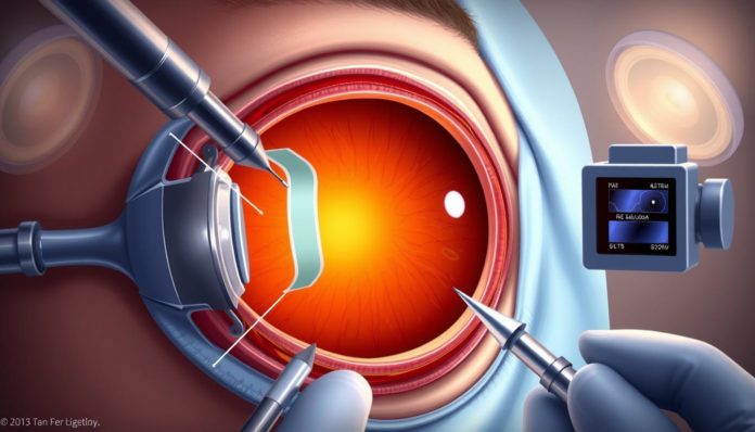
The surgery starts with giving anesthesia to make sure the patient is comfortable. Next, cryotherapy is carefully used on the retinal breaks. This freezes the area to help reattach it. Then, a silicone band is placed around the eye.
This band pushes the eye’s surface towards the detached retina. The last steps are taking out any fluid under the retina and fixing the buckle in place. This helps the retina heal the right way.
Materials Used in the Procedure
For the scleral buckling procedure, materials are picked for their lasting quality and safety with body tissue. A silicone explant is mainly used due to its flexibility and safe reaction with the body. Special sutures are used too. They are strong enough to hold the silicone in place without harming the eye.
Understanding the steps and materials in the scleral buckling procedure shows why it’s chosen for certain retinal detachments. With better surgical methods and materials, this eye surgery sees rising success rates. It gives hope to those who might lose their sight.
Scleral Buckling by a Vitreoretinal Specialist
Having eye surgery, especially for retinal detachment, means you need a true expert. A vitreoretinal specialist in ophthalmology is just that person. They are really skilled in complex eye surgeries, like scleral buckling. Their skill shows how delicate and complicated this surgery is.
Scleral buckling is a method to fix a detached retina. It uses a silicone band outside the eye. This band presses the eye’s wall onto the retina. It’s a complex procedure that requires a vitreoretinal specialist’s precise skills.
A vitreoretinal specialist’s work is vital from start to finish. They assess the situation before surgery and take care of you after. This ensures your eyesight is protected or even gets better.
Choosing a well-trained vitreoretinal specialist for scleral buckling can really improve the results. It’s essential to understand the surgery and how it affects eye health.
The use of scleral buckling by an expert is a key moment for patients with serious retinal issues. This point is where careful patient care and medical knowledge come together to help fix vision.
Recovery and Post-Surgical Care After Scleral Buckling
After a scleral buckling procedure, patients begin an important recovery phase. This phase is crucial for the success of the eye surgery. Knowing about the recovery process and sticking to post-surgical care is essential for better vision. Here, you’ll learn what the post-operative period involves and get tips for a smooth recovery.
What to Expect During Recovery
Healing from scleral buckling can take several weeks. Early on, patients might feel discomfort, experience mild pain, or notice swelling around the eye. Blurred vision is also common as the eye heals from surgery. These signs are normal, showing the eye is healing. Improvement should occur gradually.
Tips for a Successful Scleral Buckling Recovery
- Follow the Doctor’s Instructions: It’s vital to do exactly as the eye specialist says for post-surgery care. This includes taking medications as instructed to avoid infections and to control pain.
- Restrict Physical Activities: Patients should avoid strenuous activities that could stress the eye. Especially avoid lifting heavy items or bending over in the early recovery phase.
- Attend Follow-up Appointments: Going to all check-ups helps the doctor track healing. This lets the doctor adjust the treatment if needed and catch any complications early.
- Report Any Unusual Symptoms: If you notice any unusual changes like increased redness, a lot of pain, or a sudden drop in vision, call your eye surgeon right away.
Following these measures can greatly improve the recovery experience after eye surgery. Patients who stick closely to these recommendations tend to recover better. They enjoy the results of enhanced eye health sooner.
Understanding the Risks and Scleral Buckle Complications
Scleral buckling is key for fixing retinal detachment. But it comes with its risks. Scleral buckle complications and eye surgery risks need patient knowledge. We look into issues that can pop up, using advice from the Survey of Ophthalmology and Ophthalmology Times.
Post-surgery infections are rare but can happen. If not treated quickly, they could spread throughout the eye. Another major risk is a hike in eye pressure. This needs fast action to stop damage to the optic nerve.
| Complication | Potential Impact | Preventative Measures |
|---|---|---|
| Infection | Can lead to severe ocular damage if untreated. | Meticulous sterile technique during surgery and appropriate postoperative antibiotics. |
| Intraocular Pressure Increase | Potential damage to the optic nerve, possibly leading to vision loss. | Close monitoring of eye pressure post-surgery and use of pressure-lowering medications. |
| Redetachment of the Retina | May require additional surgeries, complicating recovery. | Ensure proper adherence to the postoperative regimen and regular follow-up exams. |
Bleeding inside the eye, or choroidal hemorrhage, is another concern after scleral buckling. You might see flashing lights or more floaters. Also, the retina might detach again. This needs more eye surgery, which could be more complex.
- Discuss all potential risks thoroughly with your ophthalmologist.
- Adhere strictly to post-surgical instructions to mitigate complications.
- Report any unusual symptoms immediately to your healthcare provider.
Knowing these scleral buckle complications and eye surgery risks is crucial. If you’re planning to have scleral buckling, prepare well. Understand what the surgery involves. Taking charge of your care after the surgery is essential for good results in ophthalmology.
Assessing the Success Rate of Scleral Buckling
Scleral buckling stands out as a key surgery for retinal detachment. It boasts a high success rate. This process is vital for getting the retina back in place, which helps improve vision in those with this serious eye issue.
The success of scleral buckling can vary based on different factors. These include the type of retinal detachment, how quickly treatment starts, and any pre-existing eye problems. Studies from the Archives of Ophthalmology and The British Journal of Ophthalmology have shown many patients enjoy long-lasting retinal reattachment and better vision after this surgery.
Prompt intervention and expert handling are key to maximizing the scleral buckling success rate, making it a cornerstone technique in the field of ophthalmology.
- High success rate in achieving retinal reattachment
- Improvement and preservation of visual acuity
- Dependence on prompt and precise procedure execution
Understanding the factors that affect scleral buckling’s success helps in setting realistic expectations. When discussing treatment options with eye doctors, this knowledge is crucial for making informed decisions about scleral buckling.
Alternative Treatments to Scleral Buckling for Retinal Detachment
In ophthalmology, it’s key to look at different retinal detachment treatments. Alternatives like pneumatic retinopexy and vitrectomy can better fit a patient’s specific needs. This is because each method suits different types of retinal detachments.
Pneumatic retinopexy works well when the retinal tear is in the eye’s upper half. It uses a gas bubble to push the retina back in place.
Meanwhile, vitrectomy removes the eye’s vitreous gel to reach the retina easier. It’s often used in complicated or severe retinal detachment cases. After the gel is out, a saline solution or gas bubble replaces it. This helps the retina heal properly.
| Treatment | Description | Typical Use Case |
|---|---|---|
| Pneumatic Retinopexy | Injection of a gas bubble into the eye. | Upper-half retinal detachments. |
| Vitrectomy | Removal of vitreous gel, replaced by saline or gas. | Complex or severe retinal detachments. |
These other retinal detachment treatments are huge in today’s ophthalmology. They give more tailored options to each patient’s case. This could lead to better healing and faster recovery times.
Long-Term Outlook After Scleral Buckling
After scleral buckling, it’s vital to manage patient care for lasting good results. Retina surgery follow-up and check-ups help keep patients’ eye health at its best after surgery. Catching problems early and monitoring them closely makes the surgery more successful.
Impact on Visual Acuity
Stable or better visual acuity shows the surgery went well. Regular eye checks are essential. They spot changes that could hurt vision later. This early detection is key to stopping vision problems before they get worse.
Monitoring for Long-term Complications
To avoid long-term problems, patients need routine detailed eye checks. Complications like cataracts, glaucoma, and retinal detachment require close watch. Finding and treating these issues early keeps them from harming vision for good.
Lifestyle Adjustments and Visual Aids Post-Scleral Buckling
Patients often need to change their lifestyle after scleral buckling. This helps keep the surgery results good and protects their eyes. It’s important to change daily activities and surroundings. This avoids problems and helps vision with the right visual aids.
Knowing why these lifestyle changes matter is key. They help keep the eyes healthy after surgery. This means doing things differently every day, both in small and big ways.
- Avoiding hard physical activities that could raise eye pressure or add strain.
- Making changes at work and at home to see better and feel more comfortable.
- Seeing eye care experts often to check on healing and change visual aids if needed.
Using visual aids is crucial for better vision after surgery. These aids can be special glasses or software that makes text bigger or changes how screens look. They meet each person’s needs and visual issues.
| Type of Visual Aid | Purpose | Impact on Lifestyle |
|---|---|---|
| Magnifying glasses | To make nearby objects bigger for clear vision | Helps with reading and small tasks like sewing |
| Anti-glare screens | To cut down on screen glare and reduce eye tiredness | Makes using computers for a long time more comfortable |
| High-contrast keyboards | Makes typing and using computers easier | Perfect for those who don’t see as well |
It’s vital to focus on these changes. Working with eye health experts, learning about eye care, and using the right visual aids are important. These steps make the surgery more successful. They also make life better by managing lifestyles well and using helpful visual aids.
Advancements in Scleral Buckling and Retinal Surgery Technologies
The field of ophthalmology research is always growing. New retinal surgery innovations are being developed. These advancements make surgeries like scleral buckling more precise and improve patient outcomes.
Recently, scleral buckling technology has seen big improvements in materials and methods. New biocompatible materials for buckles cause less irritation. They also work better with the body. Plus, newer imaging technologies give surgeons a clearer view of the retina. This is very important for reattaching the retina correctly.
Ongoing advancements in surgical techniques and better understanding of the ocular anatomy have significantly pushed the boundaries of what is possible in retinal surgeries.
One major improvement in scleral buckling technology is the use of minimally invasive techniques. These methods are less traumatic for patients. They also shorten surgery time and help patients recover faster.
- Enhanced imaging techniques for precise diagnostics
- Improved surgical materials that integrate seamlessly with biological tissues
- Minimally invasive approaches to reduce recovery times and increase the comfort of patients
Staying updated with the newest ophthalmology research is crucial for medical professionals. Adapting to new retinal surgery innovations is key to improving care and patient results.
The evolution of technology in ophthalmology highlights the importance of integrating research into clinical practice. Future breakthroughs in retinal surgery will likely introduce even more advanced techniques. These promise even better outcomes for patients with retinal issues.
Scleral Buckling Success Stories and Patient Testimonials
In the world of eye care, many stories showcase the success of scleral buckling. These stories include patients getting their sight back and feeling like themselves again. They share not just the success of the surgery but also how it changes their feelings and thoughts.
A look at these stories shows how important scleral buckling can be. For instance, a teacher can see clearly again to teach, or an artist can notice the fine details of color and light. These victories reflect how patients can enjoy their everyday life and hobbies once more.
These stories also tell us about the gratitude patients feel for their doctors. They talk about the journey from being diagnosed to getting better after surgery. For those facing eye surgery, these stories are a powerful message of hope. They show the joy and clear vision that can come from scleral buckling surgery.
FAQ
What exactly is retinal detachment and why is it considered a serious condition?
Retinal detachment happens when the retina separates from its supportive tissue. This leads to loss of vision. It can be permanent if not treated quickly. The retina is key for seeing, so its detachment is a major issue that needs fast medical help.
How does scleral buckling work to treat retinal detachment?
Scleral buckling puts a silicone band around the eye. This band pushes the eye’s wall inward. It helps the retina reattach to the eye’s back. The procedure often includes cryotherapy to fix retinal tears.
What determines if scleral buckling is necessary?
A thorough eye exam by a vitreoretinal specialist decides the need for scleral buckling. They look at how severe, where, and how long the detachment is. They also consider other eye problems.
What types of retinal detachments are best treated with scleral buckling?
Scleral buckling works best for rhegmatogenous retinal detachments. These are caused by tears or breaks in the retina. It can also help with some tractional retinal detachments, which happen due to scar tissue.
What should I do to prepare for scleral buckling surgery?
To prepare, you might need lab tests and a physical exam. You should stop taking some medications. Tell your ophthalmologist about your health history, current drugs, and allergies. This ensures you’re ready for the operation.
Can you explain the step-by-step process of a scleral buckling procedure?
The procedure involves giving anesthesia first. Then, cryotherapy is applied to retinal tears. A silicone band is put around the eye. Subretinal fluid is drained. Finally, the buckle is secured with sutures.
What materials are used during a scleral buckling surgery?
In the surgery, a silicone band or sponge and sutures are used. The sutures attach the implant onto the sclera, which is the eye’s white part.
Why should a vitreoretinal specialist perform scleral buckling?
A vitreoretinal specialist is trained for retinal and vitreous issues. Their skill ensures accurate diagnosis, expert surgery, and the best care in scleral buckling.
What can I expect during recovery from scleral buckling?
Recovery might bring discomfort, swelling, and blurry vision for a while. It’s important to limit activities and follow the surgeon’s care instructions for healing.
What risks and complications can scleral buckling entail?
Though usually safe, the surgery can have risks like infection, pressure increase, bleeding, or the detachment happening again. It’s key to talk about these risks with your doctor.
How successful is scleral buckling in treating retinal detachment?
Scleral buckling often works well. Many get their retinal reattached and see better. How fast you get treated and other health issues can change the results.
Are there alternatives to scleral buckling for retinal detachment?
Yes, other options are pneumatic retinopexy and vitrectomy. Pneumatic retinopexy puts a gas bubble in the eye. Vitrectomy replaces the vitreous with saline or gas.
What’s the long-term outlook for patients after scleral buckling?
Most often, patients do well over time. Many keep or get back good vision. Yet, it’s vital to check for issues like cataracts or detachment coming back with regular eye exams.
Will I need to make any lifestyle changes or use visual aids after scleral buckling?
After surgery, changing some daily habits might be needed. You might use tools to help you see better. Changing your living or work space and staying away from rough activities are good ideas.
How has scleral buckling advanced with new technologies?
Technology is making scleral buckling and retinal surgeries better and safer. New imaging tools, surgical devices, and materials for buckles are examples of these improvements.
Where can I find testimonials and success stories about scleral buckling?
You can find stories about scleral buckling in medical journals, on the web, and sometimes from eye doctors’ offices. These stories give insights into others’ experiences with the procedure.


