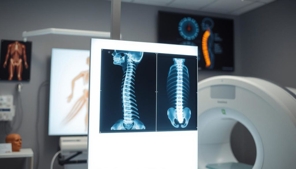
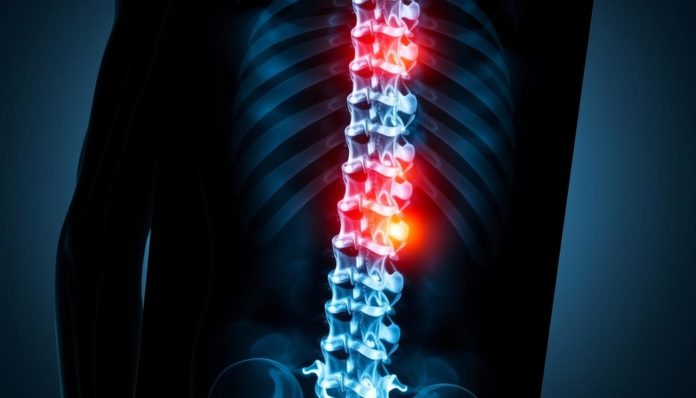
Did you know that over 80% of adults will deal with back pain? However, not all of them need a spinal X-ray. Knowing when you really need this test is key for spinal health and back pain relief.
Lower back pain usually doesn’t need an X-ray. But in certain cases, it’s crucial for making a tailored treatment plan. If you have serious symptoms or pain that won’t go away, an X-ray might be needed despite the radiation risk.
This guide will tell you more about spinal X-rays. You’ll learn when they’re necessary and their role in diagnosing issues. Keep reading for valuable information!
When is a Spinal X-Ray Needed for Back Pain?
Knowing when to get a spinal X-ray is key to treat back issues right. They are often needed when severe symptoms, called red flags, show up. These symptoms suggest there could be serious problems.

Understanding Red Flags
Red flags include things like not knowing why you’re losing weight, having had a bad injury, a history of cancer, fever, or serious loss of nerve function. When these signs are noticed, radiography for spinal issues is crucial. It helps check for major health issues like cancer or infections.
Persistent Pain Despite Treatment
If back pain doesn’t improve with usual treatments like chiropractic, massage, or physiotherapy, an diagnostic imaging for back pain might be needed. Ongoing pain, even after many treatments, could point to a missed red flag or a problem that might need surgery.
Clinicians follow proven guidelines to know when imaging is needed. This approach protects patients from unnecessary X-rays. It also helps find the exact cause of the pain efficiently.
How Spinal Imaging Helps in Diagnosing Issues
Learning about spinal imaging can make the diagnosis process for back pain clearer. This tool gives doctors a detailed view of the spine. It shows issues not seen with just a physical exam.

If you have ongoing back pain, an X-ray for spine health is key. It helps find the cause by showing fractures, infections, and more.
Different diagnostic radiology for back pain methods offer unique insights. MRI, CT scans, and X-rays each have their role. They all help doctors fully understand spine problems.
| Spinal Imaging Type | Specific Use | Benefits |
|---|---|---|
| X-Ray | Detect structural abnormalities and fractures | Quick, non-invasive, accessible |
| MRI | Visualize soft tissues, such as muscles and discs | Detailed imagery, non-ionizing radiation |
| CT Scan | Provide detailed cross-sectional images | High-resolution, good for detecting bone issues |
Using the right diagnostic radiology for back pain is crucial. It identifies spine problems and leads to effective treatment. Spinal imaging is key for better spine health, whether for simple or complex issues.
Types of Diagnostic Imaging for Back Pain
Doctors use several imaging techniques to diagnose back pain. Each method provides different details about the spine’s health. Here are the most popular types.
X-Rays
X-rays are usually the first step in checking for back issues. They quickly show us the condition of the bones. Doctors may recommend a lumbar spine X-ray, cervical spine X-ray, or thoracic spine imaging. These images help spot fractures, dislocations, or signs of wearing down.
MRI and CT Scans
An MRI for back pain is great for a closer look at soft tissues. It shows the intervertebral discs, nerves, and spinal cord clearly. This is key for identifying issues like herniated discs or spinal stenosis. On the other hand, CT scans for spinal diagnosis give detailed views of the bones. They are essential for evaluating complex fractures or unusual bone conditions.
| Imaging Type | Best For | Key Insights |
|---|---|---|
| Lumbar Spine X-Ray | Bones, Fractures | Quick and Accessible |
| Cervical Spine X-Ray | Fractures, Degenerative Changes | Efficient Initial Exam |
| Thoracic Spine Imaging | Dislocations, Abnormal Curvatures | Detailed Bone Analysis |
| MRI for Back Pain | Soft Tissues, Discs, Nerves | Detailed Soft Tissue Visualization |
| CT Scans for Spinal Diagnosis | Complex Fractures, Bone Structures | High-Resolution Bone Images |
Benefits and Risks of Spinal X-Rays
Spinal X-rays play a key role in diagnosing and treating back pain. They help doctors and patients understand what’s going on. But, it’s also important to know about the drawbacks.
Benefits
The benefits of spinal X-rays are significant. These tests quickly show the spine’s bones, spotting fractures or diseases. Doctors can see the spine’s alignment, finding problems and planning treatments.
Spinal X-rays are fast and easy for patients, making the process smooth.
Risks Involved
Despite their value, spinal X-rays have downsides. The main issue is radiation exposure. Even if it’s low, it adds up with each X-ray. Also, they can find unexpected abnormalities. This might cause anxiety and lead to more tests or treatments that aren’t needed.
| Benefits | Risks |
|---|---|
| Quick diagnosis | Exposure to radiation |
| Detailed bone structure | Potential for incidental findings |
| Non-invasive procedure | Possible patient anxiety |
Weighing the benefits of spinal X-rays against the risks of spinal radiography helps in making informed decisions. This way, patients and doctors can manage back pain diagnosis and treatment more effectively.
Preparation Tips for a Spinal X-Ray
Getting ready for a spinal X-ray can ease your worries. This safe test is often used to check the spine’s structure and find different health issues.
What to Expect During the Procedure
A healthcare worker will help you through the X-ray when you get there. Remember these important points:
- Wearing a hospital gown might be needed to avoid clothes affecting the images.
- Remove all metal items like jewelry since they can blur the X-ray pictures.
- The test is quick, only taking a few minutes, but you must not move to capture clear images.
Knowing what will happen during a spinal X-ray procedure can lessen your worry.
Pre-Procedure Guidelines
It’s crucial to follow directions before your spinal X-ray for accurate images. Before your test, your doctor might say:
- Tell the tech if there’s a chance you could be pregnant, as X-rays can risk an unborn child.
- Not to take certain meds or eat specific foods, as instructed, to ensure clear X-rays.
- To arrive early to handle paperwork and be ready for the test.
By preparing well for a spinal X-ray, you help make sure the test is successful and the images are clear.
Understanding Results of a Spinal X-Ray
When you get a spinal X-ray, you might see several common results. Knowing what these mean is key for checking spine health. It helps make sure the right treatment is planned.
Common Findings
Spinal X-rays can show different things such as:
- Vertebral fractures: These happen from injury or osteoporosis. They tell us how strong the spine is.
- Osteoarthritis: This shows wear and tear in the spine. It can lead to long-term pain.
- Spinal misalignments: Unusual shapes or shifting in the spine can affect its overall health.
Interpreting Results with Your Doctor
Looking at spinal X-rays with a doctor is important. It helps make sure the analysis of spine health is right. Talking about what’s found with your doctor considers small or age-linked changes. It also deals with serious issues fast. Working with your doctor, you can make a treatment plan that fits you.
Alternative Methods for Back Pain Diagnosis
Diagnosing back pain often requires methods beyond just imaging. Doctors perform thorough physical exams to check a patient’s motion and strength. Along with detailed history, these steps point out potential problems.
Also, doctors watch how patients move and do activities through functional assessments. This method is helpful when there’s no need for an imaging scan. It’s used unless there are signs of a serious condition.
For more on alternative back pain evaluation methods, look at trusted sources. The Mayo Clinic offers valuable information on diagnosing and treating back pain. These resources explain using non-imaging techniques to create personalized treatment plans.
Doctors use physical exams, patient history, and functional assessments to treat back pain wisely. This method avoids unnecessary X-rays and focuses on improving health with less risk. It’s a more patient-centered way to handle back pain.
Spinal X-Ray vs. Lumbar Spine X-Ray
The main difference is in the area they look at. A spinal X-ray checks the cervical, thoracic, and lumbar areas. But, a lumbar spine X-ray zooms in on the lower back.
Differences Between Procedures
It’s crucial to know the differences between spinal and lumbar spine X-rays for choosing the right one. A spinal X-ray covers a wide area. It can spot problems across the spine. But, a lumbar spine X-ray focuses on just the lower back.
Specific Uses and Benefits
A lumbar spine X-ray is great for identifying lower back issues, like disk problems or arthritis. It goes into detail where a general spinal X-ray might not see these issues. But when the pain’s source isn’t clear, a spinal X-ray is better. It checks multiple spine areas for widespread conditions.
Choosing between them hinges on what problem is suspected. This overview helps show their uses:
| X-Ray Type | Primary Focus | Best Used For |
|---|---|---|
| Spinal X-Ray | Cervical, Thoracic, Lumbar Regions | General Spinal Issues, Multi-Region Conditions |
| Lumbar Spine X-Ray | Lower Back | Specific Lower Back Problems, Disk Issues, Lumbar Arthritis |
How to Discuss Spinal X-Rays with Your Doctor
Talking about your spinal X-ray results with a doctor is key to understanding your health issue. Ask clear questions to know how these results affect your condition and care. This helps confirm your back pain reasons and ensures you make wise health choices.
Questions to Ask
Don’t be shy to ask your doctor for details. Start with questions like, “What do these spinal X-ray results say about my back pain?” Also, check if any immediate concerns pop up from the X-ray. Learning about these can help decide what to do next.
It’s smart to ask if the X-ray was really needed for your diagnosis. Knowing if there were other options can give valuable insight. Finally, find out how the results impact your treatment. This helps you know what lies ahead.
Importance of Second Opinions
Getting a second opinion is really helpful, especially if surgery is suggested. It’s a chance to confirm your diagnosis and look at different treatment paths. Sharing your X-ray with another expert can bring new ideas or less intense treatment options. Making sure multiple experts agree on your treatment ensures you’re on the best recovery route.
FAQ
When is a spinal X-ray needed for back pain?
A spinal X-ray might be needed for several reasons. It helps when there are severe symptoms, like major illness signs. It is also considered when long-lasting pain doesn’t improve with treatments like chiropractic sessions.
How does spinal imaging help in diagnosing back issues?
Spinal imaging reveals problems not seen in a physical exam. It can show fractures, infections, or changes from aging. This lets doctors tailor a treatment plan that works better for the patient.
What types of diagnostic imaging are used for back pain?
Different imaging types include X-rays, MRI, and CT scans. X-rays quickly show bone structure and any abnormalities. MRI provides closer looks at soft tissues, and CT scans offer detailed bone images.
What are the benefits and risks of spinal X-rays?
Spinal X-rays provide fast information on bone health. They help doctors diagnose and treat quickly. But, they do have small risks like radiation exposure and finding non-important issues that might cause worry.
How should one prepare for a spinal X-ray?
Preparing for an X-ray is simple. Patients should know it’s safe and involves removing metal items and wearing a gown. Knowing what to expect can reduce worry.
What are common findings in spinal X-rays?
Common X-ray findings include bone fractures and joint arthritis. Discussing these findings with a doctor is key. It helps make sure the diagnosis and treatment are correct, considering some findings might not be important.
Are there alternative methods for diagnosing back pain besides imaging?
Yes, other methods exist. They include examining the patient and understanding their medical history. These strategies help recommend treatments without needing imaging right away, especially when no severe symptoms are present.
What is the difference between a spinal X-ray and a lumbar spine X-ray?
A spinal X-ray looks at various spine regions. These can be the neck, mid-back, or lower back. On the other hand, a lumbar spine X-ray only looks at the lower back. The choice depends on what the doctor thinks is the problem.
How should I discuss spinal X-ray results with my doctor?
When discussing X-ray results, it’s vital to ask questions. Find out how the results impact your treatment and condition. Asking about the X-ray helps in making informed choices about care. Getting a second opinion can also help confirm the treatment path.


