Did you know over 24 million Americans over 40 have cataracts? This shows how crucial eye care is for catching eye diseases early. Ophthalmoscopy is a key part of keeping eyes healthy.
Ophthalmoscopy is a vital part of eye exams. It lets doctors see inside the eye to check everything is okay. This helps them make sure the retina, where the eye processes images, is healthy. It also spots signs of eye diseases early.
Doctors use direct or indirect ophthalmoscopy to see inside the eye. They dilate the pupils and examine under dim light for the best view. Knowing what ophthalmoscopy is helps us value eye check-ups more. Let’s dive into this important eye test.
What is Ophthalmoscopy?
Ophthalmoscopy, also known as funduscopy or retinal examination, is a key diagnostic tool in eye care. It lets eye doctors see inside the eye. This method is crucial for checking the eye’s inner parts.

Ophthalmoscopy Definition and Purpose
The ophthalmoscopy definition is about using special tools to look inside the eye. It focuses on the retina, optic disc, and blood vessels. The main goal is to spot eye diseases early and check the health of these important parts.
Ophthalmologists use this method to find problems quickly and start treatment right away.
The Structures Examined
During an ophthalmoscopy, doctors look at key areas such as:
- The retina, which takes in light and sends visual signals to the brain.
- The optic disc, where the optic nerve leaves the eye.
- Blood vessels, which show how well blood is flowing and can hint at other health issues.
Types of Ophthalmoscopy: Direct vs Indirect
There are two main types of ophthalmoscopy: direct and indirect.
- Direct ophthalmoscopy: This uses a handheld device with a light source for a close-up look at eye structures. It’s great for detailed checks of certain areas.
- Indirect ophthalmoscopy: This method uses a head-mounted device and lenses for a wider view. It shows the whole retina, making it good for full eye exams.
Learning about ophthalmoscopy helps us see its role in eye health. This diagnostic procedure is key for catching eye problems early.
The Importance of Ophthalmoscopy in Eye Care
Ophthalmoscopy is key in eye care because it helps find and diagnose many eye problems. It’s a safe way to check for diseases early and keep an eye on them. This helps keep eyes healthy.
Screening for Eye Diseases
Using ophthalmoscopy regularly can spot serious eye diseases like glaucoma, macular degeneration, and retinal detachment. These diseases can go unnoticed until it’s too late. Early detection is key to saving sight.
Fundus examinationduring these checks lets doctors see changes in the retina. This helps spot early signs of disease.
Assessing Overall Eye Health
Ophthalmoscopy does more than just find diseases. It’s crucial for checking overall eye health. By looking at the fundus, doctors can spot issues that might need extra care or watching.
This detailed check is a big part of regular eye health checks. It helps catch problems early, so they can be fixed quickly.
Conditions Diagnosed through Ophthalmoscopy
Ophthalmoscopy can spot more than just common eye diseases. It can find serious issues like CMV retinitis and melanoma. It also shows signs of other health problems, like diabetes and hypertension, in the eyes.

Preparing for an Ophthalmoscopy
Getting ready for an eye examination like an ophthalmoscopy is important. You need to know what to do and what to expect. Here are some tips to help you prepare:
- Dilating the Pupils: The doctor may use special eye drops to make your pupils bigger. This lets them see the inside of your eye better.
- Vision and Light Sensitivity: Your vision might get blurry, and you could feel more sensitive to light. Bringing sunglasses with you can make you more comfortable.
Talking about your health with your eye doctor is key for good eye care:
- Tell them if you’re allergic to any medicines.
- Give them a list of the medicines you’re taking now.
- Share if your family has glaucoma or other eye problems.
By doing these things, you help make sure your eye examination is thorough and helpful. This is good for your eye health.
What to Expect During the Exam
Getting ready for an ophthalmoscopy can ease your worries. This eye exam closely looks at the retina and optic nerve. It’s key to understanding your eye health.
The Procedure Step-by-Step
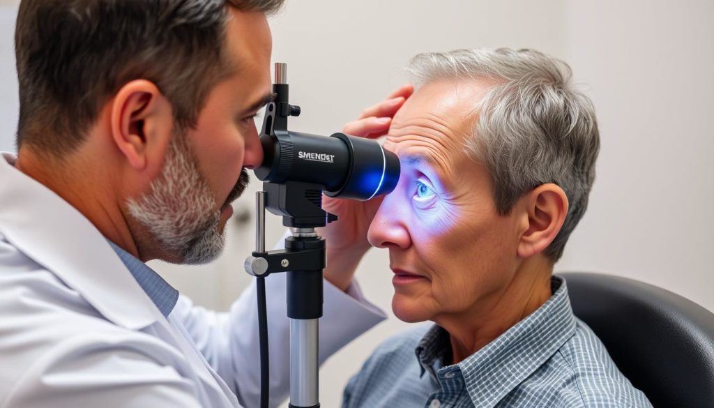
You’ll sit in a dark room during the exam. The doctor will use an ophthalmoscope to shine a light into your eye. This lets them see the inside of your eye clearly. The whole process is fast and shouldn’t hurt, but some might find the light too bright.
Types of Examinations: Direct, Indirect, Slit-lamp
There are three main ways to do ophthalmoscopy: direct, indirect, and slit-lamp. Each has its own way of looking at the eye:
- Direct Ophthalmoscopy: Uses a special tool to see the retina and optic nerve up close.
- Indirect Ophthalmoscopy: A special light and lens give a wide view of the retina. It’s great for seeing the edges of the retina.
- Slit-lamp Examination: This combines a microscope with a special lens for a super close look at the retina and optic nerve. It helps spot tiny problems.
| Type of Examination | Equipment Used | Main Benefit |
|---|---|---|
| Direct Ophthalmoscopy | Handheld ophthalmoscope | Detailed view of retina and optic nerve |
| Indirect Ophthalmoscopy | Head-mounted light and handheld lens | Wide view of the retina |
| Slit-lamp Examination | Slit-lamp microscope with special lens | Highly magnified views |
Thanks to new retinal imaging tech, these ophthalmoscopy types are now more accurate. Knowing about them can make your eye exam less scary.
Ophthalmoscopy Definition: Key Terms and Concepts
Learning about ophthalmoscopy means getting to know important terms and concepts. This includes the ophthalmoscopy definition, how a fundus examination works, and the role of retinal imaging.
Fundus, Retina, and Optic Disc
The “fundus” is the back part of the eye seen during an exam. It has important parts like the retina and the optic disc. The retina takes in visual images and sends them to the brain for processing. The optic disc, also called the blind spot, is where the optic nerve leaves the eye. This is a key part of the ophthalmoscopy definition.
Understanding Retinal Imaging and Fundus Examination
Fundus examination is vital for a full view of the eye’s inside. It helps spot eye problems. Retinal imaging is a big part of this, making detailed retina pictures. These pictures help doctors diagnose and treat eye diseases better. This imaging is key in today’s eye care, making sure exams are thorough and precise.
Risks and Side Effects of Ophthalmoscopy
Ophthalmoscopy is a key diagnostic procedure in eye care. It can sometimes cause discomfort and side effects. Knowing these risks helps patients get ready for their eye examination.
Common Discomforts
During ophthalmoscopy, some discomfort is normal. Patients might feel stinging from the dilation drops. The bright light used can also cause afterimages that may last a bit after the exam.
Potential Adverse Reactions
Some patients may have rare reactions to the eye drops used. These can include flushing, dizziness, or even severe glaucoma. It’s important for patients to know these risks and get help right away if they happen.
| Type of Reaction | Symptoms | Frequency |
|---|---|---|
| Common Discomforts | Stinging from drops, afterimages | Common |
| Adverse Reactions | Flushing, dizziness, narrow-angle glaucoma | Rare |
How Ophthalmoscopy Contributes to Diagnosing Eye Conditions
Ophthalmoscopy is a key tool in eye care. It helps doctors see inside the eye. This is crucial for keeping eyes healthy and providing the best care.
Identification of Retinal Tears and Detachment
Doctors use ophthalmoscopy to spot retinal tears and detachments. These problems need quick action to save sight. Catching them early with retinal images helps keep eyes healthy and prevents big problems.
Detection of Glaucoma and Macular Degeneration
Ophthalmoscopy is key in finding diseases like glaucoma and macular degeneration early. These diseases can cause big vision loss if caught late. By using retinal images, doctors can watch for changes and treat these diseases better.
Recognizing Systemic Health Issues Through Eye Examination
Ophthalmoscopy can also show signs of health issues like diabetes and high blood pressure. Changes in retinal blood vessels mean these problems might be present. This makes ophthalmoscopy important for overall health and eye care.
Advances in Ophthalmoscopy Technology
Ophthalmology is seeing big changes, making ophthalmoscopy better. New eye care tech is amazing, especially in showing the eye’s inner details. Now, retinal imaging and top-notch tools change how doctors do eye exams.
Digital imaging is a big step forward. It gives clear, detailed looks at the retina and other important parts. These high-res images help doctors spot and treat eye problems early, like macular degeneration and glaucoma.
Also, better lenses make ophthalmoscopy more comfortable and useful. They give sharper views without hurting the patient. These changes make exams smoother and help doctors diagnose better.
These ongoing improvements highlight the value of ophthalmoscopy and its uses. As tech gets better, it could lead to better patient care by catching problems early. The future of eye care is looking up, thanks to these advances.
FAQ
What is ophthalmoscopy?
Ophthalmoscopy is a way for eye doctors to check the inside of your eyes. They look at the retina, optic disc, and blood vessels. This helps them see if your eyes are healthy and find any problems.
What structures are examined during ophthalmoscopy?
The eye’s retina, optic disc, and blood vessels are checked during ophthalmoscopy. These parts are key for good vision. They can show signs of eye diseases or health issues in the body.
What are the different types of ophthalmoscopy?
There are two main types: direct and indirect ophthalmoscopy. Direct uses a handheld device with a light. Indirect uses a head-mounted light and lenses for a wider view inside the eye.
Why is ophthalmoscopy important for eye care?
Ophthalmoscopy is key for finding and diagnosing eye diseases like glaucoma and retinal detachment. It checks the eye’s health and can spot signs of other health issues, such as diabetes.
How should I prepare for an ophthalmoscopy?
You might need eye drops to make your pupils bigger for the exam. Tell your eye doctor about any allergies, medicines you’re taking, and your family health history, especially glaucoma.
What can I expect during an ophthalmoscopy exam?
Your eye will be examined with a bright light during the exam. The doctor will use different methods to see inside your eye clearly. You might feel some discomfort from the light, but it won’t hurt.
What are the common risks and side effects of ophthalmoscopy?
You might feel some eye discomfort from the drops used to dilate your pupils. You could also see afterimages from the light. Rarely, some people have serious reactions to the drops, like dizziness or glaucoma.
Which eye conditions can be diagnosed through ophthalmoscopy?
Ophthalmoscopy can spot retinal tears, detachment, glaucoma, and other eye problems. It also finds signs of diseases like diabetes that affect the eye’s blood vessels.
How has technology improved ophthalmoscopy?
New technology has made ophthalmoscopy better. Digital retinal imaging and improved lenses give clearer images. This makes exams more comfortable and helps doctors diagnose eye health issues more accurately.


