Welcome to a deep dive into Vitreomacular Adhesion (VMA), a key condition to know for preventing vision loss. It draws a lot of focus in ophthalmology due to its effect on how well we see. Here, we start our journey to understand VMA, from spotting it to knowing why seeing a retina expert is crucial. This chat is your first step towards guarding against the impacts of this eye issue.
Getting the right call on VMA by a retinal specialist is crucial for stopping serious vision problems. In our talk, we’ll cover living with VMA and the latest in ophthalmology that helps manage it. With our guide, you’ll gain insights that not only open your eyes but can also save your sight. Stay with us as we unpack VMA’s complexity, equipping you with life-changing knowledge.
What is Vitreomacular Adhesion?
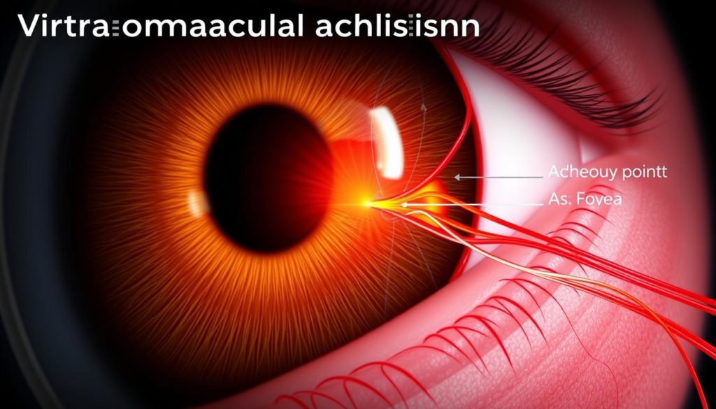
Vitreomacular adhesion (VMA) happens when the eye’s clear gel, the vitreous, sticks too much to the macula. The macula lets us see details clearly. If the vitreous pulls on the macula, it can harm our vision. VMA is known for causing eye floaters. These are the little shapes that drift in our line of sight.
VMA occurs when the vitreous, which should lightly touch the retina, doesn’t detach properly as we age. Normally, this doesn’t cause problems. But if it sticks, VMA happens. Eye floaters then appear because of fibers in the vitreous. They cast shadows on the retina.

About 2% of people over 50 get VMA, making it a common issue. While VMA can cause some discomfort and floaters, not everyone will face worse problems. Severe issues include macular holes and loss of vision. But these are not as common.
| Age Group | Prevalence of VMA | Common Symptoms |
|---|---|---|
| 50-59 | 1.5% | Minor Floaters |
| 60-69 | 2.1% | Floaters, Occasional Visual Disturbance |
| 70+ | 2.4% | Floaters, Visual Disturbance, Potential Vision Loss |
Knowing how the vitreous and macula work together is key to handling VMA. If you’re older or see floaters, get your eyes checked. Catching VMA early helps keep eyes healthy and vision clear. It shows why we all need to be aware and take care of our eyes.
Causes and Risk Factors for Vitreomacular Adhesion
Knowing what leads to Vitreomacular Adhesion (VMA) is key for catching it early and managing it well. Age, high myopia, and previous eye surgeries are major factors. They push towards issues like vitreomacular traction and even retinal detachment.
Age-Related Changes in the Eye
As we get older, our eye’s vitreous gel changes. This gel helps keep the eye’s shape but starts to liquefy and shrink with age. This shrinking pulls on the macula, risking vitreomacular traction. It can pull the macular tissue, disturb vision, and might lead to serious problems like retinal detachment.
High Myopia and Its Implications
People with high myopia face a higher VMA risk. Their elongated eyeballs stretch and thin the retina. This makes the vitreous detach unevenly, which raises the chances of vitreomacular traction. It could even result in retinal detachment.
Previous Ocular Surgeries and VMA
If you’ve had eye surgery before, like for cataracts, you might be more likely to get VMA. Such surgeries change the vitreous and eye’s inner structures. They can lead to vitreomacular traction complications. Each operation might shift the vitreous’ position or change its consistency, upping the chance for retinal detachment.

Identifying Symptoms of Vitreomacular Adhesion
Vitreomacular adhesion (VMA) symptoms differ a lot from one person to another. They can range from barely noticeable to very serious. Catching these signs early is key to manage them well.
At first, the symptoms might not seem like a big deal. Yet, they could get worse over time. They might even lead to a macular hole. Here are the common symptoms of VMA:
- Distortion of vision – where straight lines look wavy or bent
- Difficulty reading or doing detailed tasks
- Images seem smaller or further away
- Blurriness and texture changes in central vision
These issues can make daily tasks hard. Things like reading, driving, and recognizing faces become tough. If these changes happen, seeing a doctor is crucial to avoid more vision loss.
Early intervention can prevent conditions like macular holes, which are often a severe complication of untreated VMA.
If symptoms get worse, it may point to a macular hole. This needs quick medical help:
- An increase in floaters, small dots or lines that float in your vision.
- Seeing flashes of light.
- Obvious loss of vision in one or both eyes.
Here’s a chart showing how VMA symptoms can get worse over time:
| Stage | Common Symptoms | Risk of Progressing to Macular Hole |
|---|---|---|
| Early | Visual distortion, difficulty with fine details | Low to moderate |
| Moderate | Blurred central vision, perceived changes in object size | Moderate |
| Advanced | Significant loss of central vision, flashes, floaters | High |
It’s vital to spot these symptoms early. Knowing they can lead to serious problems is important. Regular doctor visits are crucial. They help manage symptoms well and prevent major vision loss.
Vitreomacular Adhesion and its Potential Complications
Vitreomacular adhesion (VMA) can seriously affect your eye. It might make you need surgery. Knowing about it helps patients and doctors choose the right treatment.
Progression to Macular Hole
VMA can pull at your macula, creating a hole. This hole can blur and distort your vision. Surgery is often needed to fix it.
Understanding Vitreomacular Traction
Vitreomacular traction (VMT) happens when the vitreous humor doesn’t detach from the retina as it should. It pulls on the retina, causing swelling and distorted vision. This might lead to conditions that need surgery to fix.
Retinal Detachment Risks
Severe VMA tractions can tear the retina. Ignored, it might lead to the retina detaching. This is urgent and requires surgery to save your sight.
| Condition | Symptoms | Recommended Intervention |
|---|---|---|
| Macular Hole | Blurring and Distortion | Possible vitrectomy surgery |
| Vitreomacular Traction | Visual Distortion and Swelling | Monitoring, possibly surgical intervention |
| Retinal Detachment | Sudden Severe Visual Changes | Urgent vitrectomy surgery |
Diagnosis Procedures for Vitreomacular Adhesion
Finding out if someone has vitreomacular adhesion (VMA) is very important. It’s mainly done by a retinal specialist in the eye care field. This part tells about the many high-tech tools and methods used to correctly find VMA.
Optical Coherence Tomography (OCT) is a key method for getting detailed pictures of the retina. It lets eye doctors see the vitreomacular interface clearly. This safe way is crucial for diagnosing and keeping track of how VMA changes.
- Fundus photography: This takes clear pictures of the retina. It’s used for keeping records and watching how the retina changes over time.
- Fluorescein angiography: This method uses a special dye to highlight blood vessels. It can show any unusual activity or damage in the retina’s blood vessels.
- B-scan ultrasonography: This is helpful when OCT can’t get a good look due to blockages. It shows a picture of the inside of the eye.
To help you understand better, here’s a comparison of these imaging methods:
| Technique | Use in VMA Diagnosis | Benefits |
|---|---|---|
| Optical Coherence Tomography (OCT) | Standard imaging for diagnosing VMA | High-resolution images, non-invasive, no radiation exposure |
| B-scan ultrasonography | Used when OCT is not viable | Can image through opacities, provides view of the complete eye structure |
| Fundus photography | Helps document and monitor retinal status | Effective in tracking disease progression |
| Fluorescein angiography | Assesses blood vessel health | Identifies leakage or blockage in blood vessels |
Getting diagnosed early and correctly by an experienced eye doctor is key to managing VMA well. It also helps prevent worse eye problems. These tools give a full check-up and help choose the best treatment for each person.
Treatment Options: From Observation to Surgery
Treating Vitreomacular Adhesion (VMA) depends on the condition’s severity and what the patient needs. Options range from simply watching the condition to using advanced surgery. The goal is to both keep and improve how well you can see.
Monitor or Wait: Observing VMA
Many times, doctors suggest just watching the condition, especially if it’s not really affecting your life. This means going to the eye doctor often. They check if the condition is getting worse or causing other issues.
Pharmacological Interventions and Injections
When more action is needed, pharmacological interventions usually come first. These might be special drugs to reduce symptoms or stop VMA from getting worse. Injections in the eye can help too. They work by easing the vitreomacular traction, all without having to do surgery.
Vitrectomy Surgery and Recovery
In severe cases where other treatments don’t work, vitrectomy surgery might be needed. This operation takes out the vitreous gel to prevent it from pulling on the retina. This is important since that pulling can cause holes or tears. After surgery, watching how you recover is crucial. It helps to prevent any problems and ensures your vision gets as good as possible.
Understanding the Vitrectomy Procedure in Detail
Vitrectomy surgery tackles eye conditions like vitreomacular adhesion (VMA) to prevent vision loss. It involves removing the vitreous gel from the eye to maintain retinal health. This discussion explores the surgery’s goals and what recovery looks like.
A vitrectomy becomes necessary when other treatments can’t stop diseases affecting the retina. This procedure treats current problems and prevents worse issues, like losing sight.
- It removes the vitreous gel for better surgical retina access.
- It addresses retinal tears or detachment to avoid blindness.
- It takes out scar tissue that could damage the retina.
Vitrectomy surgery demands high skills and advanced technology. Ongoing advancements in tools and methods help surgeons protect delicate retinal tissue and reduce surgery trauma.
Modern vitrectomy methods lead to better vision and fewer problems afterward.
Doctors evaluate patients carefully to see if a vitrectomy is right for them. They look at how bad the vision problem is, the condition of the eye, and the patient’s general health.
It’s important to know the possible upsides and downsides of this surgery. While vitrectomy surgery often works well, it can sometimes lead to infections, bleeding, or higher eye pressure. These issues can make vision worse if they’re not treated quickly.
Choosing to have a vitrectomy can be key to keeping good vision and life quality for those with severe retinal issues. With good information and a skilled doctor, patients can make informed decisions about their treatment.
Post-Surgical Care and Managing Expectations
After vitrectomy surgery, the road to better vision includes important steps. You need to take care of your eye, work on getting your sight back, and see your eye doctor often. Handling things well and having realistic hopes are vital for good results.
Immediate Post-Op Care
Care starts right after the surgery. You must follow a strict plan that includes taking medicine to stop infection and reduce swelling. It’s important to keep the eye safe and stay away from hard activities or places that could bother the eye. Seeing the doctor often after surgery helps check on healing and find any problems early.
Long-term Vision Rehabilitation
Getting your vision back is key to recovery, especially if your sight was really bad before the surgery. This could mean doing special eye exercises, using certain tools, or going to therapy programs. Being patient and steadfast is important, as it may take months to see the best results.
When to Contact Your Retinal Specialist
Keeping in touch with your eye doctor is crucial while you recover. You should tell them right away if your vision changes suddenly, you feel more pain, or see signs of problems. Going for regular check-ups is essential, but knowing when to get urgent help is necessary for the best eye care.
Following these steps and working closely with your doctors can help make your recovery smoother. It can also help you get better outcomes after your eye surgery.
The Role of Retinal Specialists in Managing VMA
In the field of ophthalmology, retinal specialists play a key role in handling vitreomacular adhesion (VMA). They’re experts not just in diagnosing but also in the full care needed to address this eye issue.
Retinal specialists use high-end tools to pinpoint VMA, crucial for the right treatment. This careful method helps avoid worse issues like vision loss or detachment.
They create personalized plans for VMA patients, from monitoring to possible surgery. They rely on the newest ophthalmology research and tech, aiming for the best care.
- Regular monitoring with high-quality imaging.
- Guiding patients on disease progress.
- Performing surgeries like vitrectomy if needed.
Seeing a retinal specialist often is vital for those with VMA. These specialists do more than treat. They also teach and strengthen patients in handling their condition.
Retinal specialists are essential in ophthalmology, especially for those dealing with VMA. Their knowledge and skills are critical in the broad scope of eye health.
Vitreomacular Adhesion and Lifestyle Considerations
Living with vitreomacular adhesion (VMA) means making some changes to your life. This is to handle changes in how well you can see and to keep your eyes healthy. Learning about these changes helps you deal with VMA and improves your life.
Daily Activities and Visual Demands
Those with VMA need to look closely at their daily visual tasks. Reading, driving, or using computers might get tougher. You might need better lighting, tools to make things look bigger, or breaks to rest your eyes.
Nutrition and Eye Health
Eating right is key in managing VMA. Vitamins C and E, zinc, lutein, and omega-3s are good for your eyes. They might slow down the symptoms. Try to eat lots of fruits, leafy greens, fish, and nuts every day.
Protecting Your Eyes Post-Diagnosis
After finding out you have VMA, it’s important to protect your eyes. Wear sunglasses against UV rays and safety glasses for risky activities. Also, see an eye doctor often to check on VMA.
| Activity | Modification | Benefit |
|---|---|---|
| Reading | Use magnifying aids | Reduce strain and improve clarity |
| Computer Work | Follow the 20-20-20 rule | Decrease fatigue and dry eyes |
| Outdoor Activities | Wear UV protection sunglasses | Protect from harmful UV rays |
Adjusting to new visual demands is critical in handling VMA well. Simple but smart changes in daily life, focusing on what you eat, and careful eye protection make a big difference. They help you manage VMA and live a better life.
Patient Stories: Living with Vitreomacular Adhesion
Learning about vitreomacular adhesion through patient stories gives us key insights. These stories do more than talk about what the condition is. They show how deeply it affects people’s lives. We share stories from those who have faced the ups and downs of vitreomacular adhesion.
It was overwhelming at first, learning about my diagnosis. But hearing how others navigated their journey gave me hope and a path forward.
These stories highlight the hurdles and the strength needed to live well with this condition. Even with fear and doubt, support and good medical advice bring hope. This helps people find ways to adjust and cope.
- Understanding and accepting the diagnosis
- Finding the right specialist and treatment plan
- Adapting lifestyle and daily activities
- Connecting with support groups and other patients
Patient stories build a sense of community. They also help raise awareness, leading to better care and new treatments for vitreomacular adhesion.
More stories mean a richer understanding. Every story adds to our collective knowledge. Through these accounts, we see human resilience in the face of adversity. These stories emphasize the need for kindness, more studies, and customized care plans.
Advancements in Ophthalmology for VMA Treatment
Recent advancements in ophthalmology have transformed VMA treatment. With ongoing future research, new treatment options are emerging. These developments offer hope and better outcomes to those suffering from VMA.
Emerging Medical Therapies
Groundbreaking work has led to new medical therapies for VMA. These include enzymatic and pharmacologic approaches that avoid surgery. They offer a non-invasive way to treat the condition, giving patients better options.
Technological Innovations in Surgery
Technological advances have made surgery for VMA safer and more precise. New tools and real-time imaging systems lower the risks of vitrectomy. They make these procedures much safer for patients.
Future Directions in Research and Care
Exciting research is being done in gene therapy and artificial intelligence for VMA. These efforts aim to improve diagnoses and treatments. They promise to make patient care better and more efficient.
The table below shows how new surgical methods compare to old ones. They are safer and more effective, thanks to technological progress.
| Traditional Surgery Techniques | Advanced Surgical Techniques |
|---|---|
| Manual instruments | Automated cutting tools |
| Standard visualization | Enhanced imaging systems |
| Longer recovery times | Minimized recovery periods |
This changing field dramatically improves VMA treatment and patient life quality. It marks a significant shift in eye care.
Practical Advice for Preventing Vision Loss
To keep your eyes healthy and avoid issues like vitreomacular adhesion (VMA), there are key steps to follow. One important action is to get regular eye exams. These aren’t just to update glasses or contacts; they catch early signs of VMA or other problems.
Keeping an eye on your vision health means more than just visits to the optometrist. It requires noticing changes in your vision, such as blurriness or floaters. If you see these signs, contact a retinal specialist quickly. Early detection can help manage eye conditions better, helping to save your vision.
Finally, adopting eye-friendly habits is crucial for vision loss prevention. Eat well, exercise to improve blood flow, and wear sunglasses to shield from UV rays. Prevention is key, so be proactive in protecting your valuable sense of sight.
FAQ
What exactly is Vitreomacular Adhesion, and how does it occur?
Vitreomacular Adhesion (VMA) happens when the eye’s vitreous gel sticks to the macula. This is the part of the eye that lets us see details. The gel can shrink with age and pull on the macula, possibly harming vision.
Are eye floaters related to VMA?
Yes, eye floaters can be a sign of VMA. They occur when the vitreous gel pulls away from the retina. This is part of getting older. Floaters look like small shadows or specks moving in what you see.
What causes VMA, and am I at risk?
Getting older raises the risk of VMA. It’s also more common in those with severe nearsightedness or who had eye surgery. VMA happens when the vitreous gel changes and sticks more to the retina.
How do I know if I’m experiencing symptoms of VMA?
VMA symptoms include blurry vision or seeing straight lines as wavy. Having trouble with tasks needing detailed vision can also be a sign. See a specialist if you notice changes in your vision.
What are the possible complications of untreated VMA?
Not treating VMA can lead to more serious issues like vitreomacular traction or a hole in the macula. Worst case, it could cause the retina to detach, which is an emergency that might result in losing vision.
What should I expect during a vitrectomy surgery?
In a vitrectomy surgery, the doctor takes out the vitreous gel. This helps remove the pull on the macula. Then, they fill the eye with saline, gas, or silicone oil. This keeps the eye’s shape and helps it heal.
Following vitrectomy surgery, how should I manage my recovery?
After surgery, you’ll use eye drops to protect against infection and swelling. You’ll need to avoid certain activities and keep seeing your specialist. They’ll give you specific recovery steps.
How do retinal specialists contribute to managing VMA?
Retinal specialists are key in managing VMA. They diagnose the condition, recommend how to watch it or treat it, and can perform surgeries to save vision.
What lifestyle changes might I need to consider after a VMA diagnosis?
After a VMA diagnosis, you may need to change some daily habits to protect your eyes. Eating well for eye health and avoiding eye strain are important. Taking care to prevent further eye damage is also key.
Are there new treatments for VMA on the horizon?
Yes, the science of eye care keeps advancing. New surgical methods and medical treatments are being developed. They aim to better treat VMA and improve patient results.
How can I proactively prevent vision loss?
To fight vision loss, see a retina expert regularly. Be on the lookout for early signs of eye problems like VMA. A healthy lifestyle that supports eye health is also vital.
What advancements have been made in vitrectomy surgery?
Vitrectomy surgery has seen big improvements. Now, there are less invasive ways to do it, better imaging for precision, and faster recoveries. This all leads to better results for those with VMA.
When should patients with VMA return to their retinal specialist?
People with VMA should stick to the check-up schedule their eye doctor recommends. If their vision suddenly gets worse, they need to see their specialist right away. This is to catch any complications early or to get more treatment if needed.


