Did you know about 70% of Pancoast tumor cases are often missed at first? This is because their signs are hard to catch. They usually don’t appear on the regular chest X-rays doctors use. Knowing the unique Pancoast Tumor Symptoms is key for catching it early and treating it well.
Pancoast tumors cause different signs than other lung cancer symptoms. Instead of coughing up blood, you might have bad shoulder and arm pain. There can also be arm weakness and Horner Syndrome. Spotting these early can really help improve the chances of getting better.
Let’s look more into these tumors, like the usual signs, how to diagnose them better, and why pictures are so important for finding them early. Begin your learning journey with us now.
What is a Pancoast Tumor?
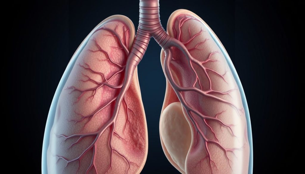
Pancoast tumors are a special kind in chest cancer because of where they grow and their effects. These tumors start at the top of the lungs, known as the “lung apex”. They usually hit the chest’s structure more than the lung itself. Their position means they can spread to nearby areas, causing certain symptoms and problems.

Pancoast Tumor Definition
Pancoast tumors belong to a group called non-small cell lung cancer. They start at the “lung apex” and often spread to close areas, like ribs, spine, and lymph nodes. They’re mainly known for affecting the chest wall and messing with nerves and blood vessels around them.
Location and Structure
A Pancoast tumor’s specific spot at the “lung apex” ties it to important chest areas. These tumors can press on or grow into “brachial nerves”. This can cause a lot of pain and even damage the nerves. The effect on “chest wall structures” can lead to shoulder pain and make your arm weak, making it harder to figure out and treat the issue.
Know the details of where Pancoast tumors are and how they’re built is key. Doctors need this info to see the complex parts where the “lung apex”, “chest wall structures”, and “brachial nerves” meet. This helps them come up with good treatment plans.
Common Pancoast Tumor Symptoms
Pancoast tumors show signs that can lead to an early catch and better treatment. It’s key to know these symptoms for quick doctor visits.

Shoulder Pain
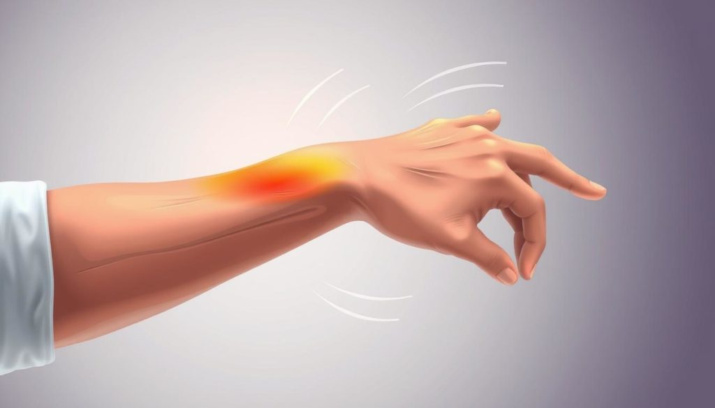
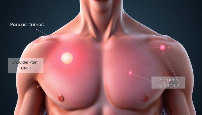
The most common sign of a Pancoast tumor is severe and constant pain in the shoulder. This pain often spreads to the arm, elbow, and fingers. It’s usually so bad that narcotic pain medications are needed.
Arm Weakness
As the tumor grows, arm weakness and muscle atrophy might happen, alongside tingling. These issues come up as the tumor pressures nearby nerves and vessels, hurting muscle action and power.
Horner Syndrome
Getting Horner Syndrome also points to a Pancoast tumor. It shows as a droopy eyelid, reduced sweating on half the face, and a smaller pupil. These signs mean the tumor is messing with nerves.
| Symptom | Description | Possible Cause |
|---|---|---|
| Shoulder Pain | Severe and constant pain that extends to the arm, elbow, and fingers. | Tumor pressing on nerves |
| Arm Weakness | Decrease in muscle strength, possibly leading to muscle atrophy. | Nerve and blood vessel compression |
| Horner Syndrome | Drooping eyelid, lack of facial sweating, pupil narrowing. | Nerve involvement |
The Role of Imaging in Diagnosing Pancoast Tumors
Imaging techniques play a key role in finding and sizing up Pancoast tumors. It’s vital to know each method’s pros and cons to fully understand this condition.
Chest X-ray Limitations
A chest X-ray is often the first step in checking for these tumors. But, it struggles to show the top part of the lung well. That’s where Pancoast tumors usually are. So, it might not always give us all the answers.
CT Scan Benefits
A CT scan gives a clearer picture than an X-ray. It shows if the tumor has spread to nearby areas like the ribs or spine. This info makes the CT scan a crucial step in figuring out and managing Pancoast tumors.
MRI Accuracy
For the most precise look, MRI is the best. It beats CT scans in showing how much the tumor has grown. This includes whether it has moved into the brachial plexus or nearby tissues. MRI’s detailed imaging is key for diagnosing correctly.
Besides these main imaging methods, arteriograms, venograms, and bronchoscopies also help with diagnosis. But, getting a tissue biopsy is vital. It’s the only way to know for sure if the tumor is cancer.
| Imaging Modality | Strengths | Limitations |
|---|---|---|
| Chest X-ray | Initial assessment, widely available | Poor visualization of lung apex |
| CT Scan | Detailed view, good for staging | Less effective than MRI for soft tissue invasion |
| MRI | Accurate in assessing tumor extent | More expensive, longer examination time |
Advanced Diagnostic Tests for Pancoast Tumors
Advanced tests are essential for diagnosing Pancoast tumors. They help catch them early and understand them better. This understanding is key for the right treatment. Let’s examine three main ways to diagnose them:
Needle Biopsy
A needle biopsy is often used to diagnose Pancoast tumors. It’s a simple method that takes tissue samples for pathology lab testing. This test is very accurate, with success rates up to 95%. It’s less invasive, making it a preferred option for many.
Video-Assisted Thoracoscopy Surgery (VATS)
VATS lets doctors see inside the chest. They make tiny chest incisions to insert a thoracoscope and tools. This method improves tumor detection and speeds up patient recovery. It’s better than older methods.
Thoracotomy
Thoracotomy is used when other tests don’t give clear answers. It’s a big chest incision to get to the lungs. It’s best for removing tumors and thorough pathology lab testing. Though more invasive, it’s effective for avoiding extra surgeries.
Stages of Pancoast Tumors
To treat Pancoast tumors right, knowing their stage is key. The TNM system helps doctors figure this out. It looks at tumor size (T), if lymph nodes are involved (N), and if it has spread (M).
Most Pancoast tumors are far along by the time they’re found, often at stages T3 or T4. At these stages, the cancer has grown into nearby areas, like the spinal cord. This makes deciding on treatment tricky.
The TNM system shows how much the cancer has spread. Symptoms like shoulder pain and weight loss mean the cancer might be advanced. It’s important to check symptoms closely.
Choosing the right treatment, such as surgery or chemotherapy, depends on the tumor’s stage. More info on symptoms and stages is available here.
Now, let’s see what each part of the TNM system tells us about Pancoast tumors:
| Stage | Tumor Size (T) | Lymph Node Involvement (N) | Metastasis (M) |
|---|---|---|---|
| T1 | Small tumor confined to lung | No lymph node involvement | No metastasis |
| T2 | Larger tumor or involving nearby structures | Possible regional lymph node involvement | No metastasis |
| T3 | Significant invasion into the chest wall | Regional lymph node involvement | No metastasis |
| T4 | Invasion into vital structures such as the spinal cord | Significant regional lymph node involvement | Possible distant metastasis |
Using the TNM system to stage Pancoast tumors is vital for the best care plan. Knowing whether a tumor is stage T3 or T4 helps understand its impact. This knowledge is crucial for planning treatment and predicting outcomes.
Risk Factors for Developing Pancoast Tumors
Knowing what increases the risk of Pancoast tumors can lead to early detection. Many factors that cause these tumors align with those for lung cancer.
Smoking and Secondary Smoke
Smoking is a top reason people get lung cancer, including Pancoast tumors. Non-smokers can also be at risk if they’re around smoke often. This is especially true for those regularly in smoky places.
Industrial and Environmental Exposure
Being around industrial toxins like chromium and nickel raises lung cancer risk. Those in industrial jobs need to watch out, as such exposure can lead to Pancoast tumors.
Asbestos and Other Carcinogens
Asbestos and similar dangers are significant risk factors too. Even less well-known substances add to the danger of lung cancer. Knowing and avoiding them can reduce risks.
| Risk Factor | Description |
|---|---|
| Smoking | Primary cause of lung cancer; significant contributor to Pancoast tumors |
| Secondary Smoke Exposure | Non-smokers exposed to smoke; increased risk |
| Industrial Carcinogens | Exposure to chromium, nickel, and others in industrial settings |
| Environmental Carcinogens | Includes asbestos; extended exposure increases risk |
Differentiating Pancoast Syndrome from Other Conditions
Diagnosing Pancoast syndrome is tough because its symptoms are similar to other health problems. Accurate diagnosis is vital for the right treatment. It’s often hard to tell it apart from muscle and bone disorders.
Musculoskeletal Disorders
Shoulder and upper arm pain is a key sign of Pancoast syndrome. But, this pain can come from other issues too. Problems like rotator cuff injuries and frozen shoulder share these symptoms. That’s why doctors need to perform detailed tests, such as imaging, to make the correct diagnosis.
Other Thoracic Neoplasms
Other chest tumors might cause symptoms like those seen in Pancoast syndrome. Conditions like mesothelioma need different treatments. Tools like x-rays and MRIs are needed for a proper diagnosis. According to research, these methods are crucial for finding the cause of symptoms and choosing the right treatment. Check out this study on Pancoast syndrome for more info.
Prognosis and Survival Rates for Pancoast Tumor Patients
Pancoast tumors are a rare lung cancer type, posing distinct challenges. Yet, medical advances have significantly improved patient outcomes. Now, survival rates for those in early stages can hit 30%-50% five years after treatment. This leap in survival shows the power of modern treatments like chemoradiotherapy and surgery.
The blend of chemoradiotherapy is key in boosting survival chances. It shrinks the tumor before surgery and attacks any remaining cancer cells. Surgical removal of the tumor is crucial. When it follows chemoradiotherapy, it raises the odds of effective treatment and longer life.
To complement treatment, it’s essential to quit smoking and avoid cancer-causing environments. Changes in lifestyle matter for patients and those at risk. With ongoing advancements in healthcare, the future for Pancoast tumor patients looks bright. Early detection and thorough treatment play pivotal roles.
FAQ
What are the early symptoms of a Pancoast tumor?
The early signs of a Pancoast tumor might not be obvious. You may feel intense shoulder pain that moves down to your arm, elbow, and fingers. As it grows, the tumor can lead to muscle weakness and loss of muscle in your arm.
Why can Pancoast tumors be difficult to diagnose?
Finding a Pancoast tumor early on is hard. They often don’t appear on regular chest X-rays. Special tests like CT scans or MRIs are needed to spot them due to their position at the lung’s top.
What is Horner Syndrome and how is it related to Pancoast tumors?
Horner Syndrome is when one eyelid droops, there’s less sweating, and the pupil is small on one side of the face. It shows up when a Pancoast tumor affects certain nerves and is a sign the tumor might be advanced.
What imaging tests are used to diagnose Pancoast tumors?
To diagnose a Pancoast tumor, doctors rely on CT scans and MRIs. These scans are better than chest X-rays for seeing the tumor’s exact location and size.
What are the benefits of a CT scan in diagnosing Pancoast tumors?
A CT scan gives clear pictures that can show if the tumor has spread to nearby areas. It’s often better than an X-ray for seeing the tumor clearly.
How accurate is an MRI in diagnosing Pancoast tumors?
An MRI is very good at showing how much the tumor has grown and if it’s moved into nearby spaces. This makes it very important for getting the right diagnosis.
What is involved in a needle biopsy for Pancoast tumors?
For a needle biopsy, a doctor takes a small piece of the tumor with a needle. This sample is then tested in a lab. This test can confirm the diagnosis in most cases.
How is Video-Assisted Thoracoscopy Surgery (VATS) used in diagnosing Pancoast tumors?
VATS is a small surgery where a camera is put into the chest through a small cut. This lets doctors see inside and find tumors without a big surgery.
What is the role of thoracotomy in diagnosing and treating Pancoast tumors?
A thoracotomy is a bigger surgery that lets doctors see and reach the lung directly. This can help in getting a biopsy or even removing the tumor, often avoiding the need for more surgeries.
What are the stages of Pancoast tumors?
Pancoast tumor stages are set by the TNM system, looking at tumor size, lymph node involvement, and whether it’s spread. They’re often found at later stages, such as T3 or T4.
What are the primary risk factors for developing Pancoast tumors?
Things that raise your risk include smoking, being around secondhand smoke, and long-term contact with industrial chemicals like chromium, nickel, and asbestos.
How can Pancoast Syndrome be differentiated from other conditions?
Telling Pancoast Syndrome apart from other issues is key. It often starts with symptoms like pain in the shoulder and arm, which could be mistaken for other health problems.
What are the survival rates for patients with Pancoast tumors?
The outlook for Pancoast tumor patients has gotten better. Now, up to 50% of people in early stages may survive for 5 years after getting treatment that combines chemotherapy, radiation, and surgery.


