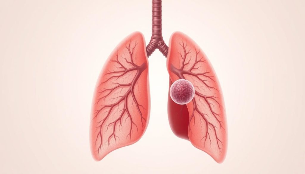
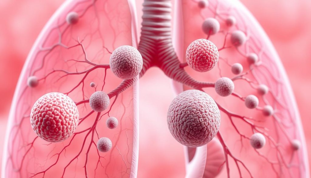
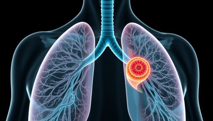
Did you know up to 90% of solitary pulmonary nodules (SPNs) are found by chance? They’re spotted during imaging for other health problems. This fact shows we need to focus on these lung spots. SPNs are small, round shadows in the lung tissue. At first, they might not seem important. However, they could be an early sign of lung cancer.
Evaluating SPNs well is key. Lung cancer is the top cause of cancer deaths in the United States. An SPN smaller than 3 cm might look harmless. But, without signs like lymphadenopathy or pneumonia, it needs a careful check. This is to ensure there’s no cancer. Knowing the risks SPNs carry helps doctors and patients act early for lung health. This could save lives.
Introduction to Solitary Pulmonary Nodule

A solitary pulmonary nodule (SPN) is often found during lung check-ups. It’s essential to know what it means and how to handle it for the best care in solitary lung nodule management.
Definition
An SPN is a small, separate lung spot under 3 cm wide. It doesn’t connect to nearby lung parts, like the outer lung layer or big airways. There’s usually no sign of lymph node issues. Still, some can be cancerous, which is why doctors must watch them closely and figure out if they’re harmful through SPN detection.
Importance of Early Detection
Spotting an SPN early is key to helping patients do well. Many times, these nodules don’t cause symptoms, so regular scans, like CTs, are crucial. Finding them early means doctors can act fast. This helps tell apart harmless from harmful nodules and decide on the right treatment plan.
Prevalence
SPNs show up in about 0.1% to 0.2% of chest X-rays and as much as 13% of CT scans. They’re more common in smokers and people who’ve had cancer before. Given how often they occur, it’s important for both doctors and patients to stay alert and follow through with strong solitary lung nodule management practices.
Common Causes of Solitary Pulmonary Nodules
Solitary Pulmonary Nodules (SPNs) can come from several sources. It’s key to know these to diagnose and treat accurately. Main causes include infections, benign growths, and cancerous tumors. They all mean different things for the patient.
Infectious Granulomas
Infectious granulomas are a top reason for SPNs. They mainly come from bacterial and fungal infections. Tuberculosis and histoplasmosis are big examples. These lead to granulomatous inflammation, causing nodules.
Benign Neoplasms

Benign tumors also cause SPNs. Hamartomas and fibromas are examples of non-cancerous lung tumors. They’re important to identify and watch. This helps avoid complications and surgeries that aren’t needed.
Malignant Neoplasms
The most concerning SPN causes are malignant neoplasms. This includes primary lung cancer and metastatic diseases. Finding these nodules early is crucial. It can save lives, as timely treatment matters a lot for lung cancer.
Symptoms Associated with Solitary Pulmonary Nodules
Most people with solitary pulmonary nodules (SPNs) don’t show symptoms. But, symptoms can occur, depending on what’s causing the SPN, like infections or cancer. Finding symptoms early is key for diagnosing and treating the condition.
Look out for these lung nodule signs and symptoms:
- Persistent cough
- Unexplained weight loss
- Chest pain or discomfort
- Shortness of breath
- Recurrent respiratory infections
These symptoms might not happen often, but they stress the need for careful checks. Without detailed tests, like chest X-rays or CT scans, it’s easy to miss the pulmonary nodule clinical presentation.
Full clinical exams and imaging matter a lot. SPNs might be nothing to worry about or could signal something severe, such as lung cancer. Finding them early is critical to treat them effectively.
Imaging Techniques for Detecting SPNs
Detecting SPNs correctly depends on using advanced imaging techniques. Doctors start with basic imaging and move to more advanced methods as needed.
Chest X-ray
The first step in checking for an SPN is a chest x-ray. While it’s fast and available everywhere, it might miss small or subtle nodules. Still, it provides early insights and can show if more tests are needed.
Computed Tomography (CT) Scan
A CT scan for lung nodule is key to checking SPNs. CT scans show detailed images, which help doctors track the nodule’s size, shape, and changes. Its accuracy is crucial for telling harmless growths from harmful ones.
Positron Emission Tomography (PET) Scan
For patients with a high cancer risk, a PET scan diagnosis offers more info. PET scans look at how the nodule behaves, providing clues about its nature. High activity on a PET scan usually means cancer, while low activity suggests it’s benign.
| Imaging Technique | Purpose | Advantages | Disadvantages |
|---|---|---|---|
| Chest X-ray | Initial screening | Quick, accessible | Low sensitivity, limited detail |
| Computed Tomography (CT) Scan | Detailed evaluation | High resolution, precise | Higher radiation dose, costlier |
| Positron Emission Tomography (PET) Scan | Further evaluation of high-risk cases | Assesses metabolic activity | Expensive, requires specific facilities |
To sum up, using SPN imaging methods like chest x-rays, CT scans, and PET scans together is key. Each one has its own strengths and weaknesses. Their use together is vital for correct diagnosis and treating SPNs well.
Risk Factors for Malignancy in SPNs
Some factors make it more likely for an SPN to be cancerous. Knowing these can help us focus on patients who need quick and thorough care.
Smoking History
Smoking is a big risk factor for lung nodules. Smokers are at a higher risk for cancerous SPNs. That’s why patients who’ve smoked a lot need careful checks.
Age and Gender
How old you are and whether you’re a man or a woman affects your SPN cancer risk. Older people and men are at a higher risk. Thus, diagnosing and managing their condition takes extra caution.
Previous Malignancies
Having had cancer before increases the risk of SPN being malignant. These patients are more likely to get cancerous lung nodules. They need detailed exams and ongoing watchfulness.
Differential Diagnosis of Solitary Pulmonary Nodules
It’s very important to know the cause of solitary pulmonary nodules (SPNs). Finding out if a nodule comes from an infection, inflammation, or birth condition helps doctors decide how to treat it.
Infectious Causes
SPNs can come from infections like tuberculosis and histoplasmosis. These infections create granulomas, looking like nodules on scans. It’s key to consider how common these diseases are in the area when checking SPNs, especially in places where they are often found
Inflammatory Causes
Rheumatoid arthritis and sarcoidosis are linked with SPNs. They cause inflammatory nodules from the immune system fighting back. Looking at the patient’s history and doing tests helps find these lung nodule causes.
Congenital Causes
Some SPNs are from conditions people are born with, like bronchogenic cysts and arteriovenous malformations. These are usually found by accident on scans. Knowing what causes these nodules early on helps manage them right.
Guidelines for SPN Follow-Up
Managing solitary pulmonary nodules (SPNs) properly requires following specific pulmonary nodule guidelines. The Fleischner Society‘s guidelines are widely used for this purpose. They propose a well-thought-out, evidence-based method for SPN follow-up.
Fleischner Society Guidelines
The Fleischner Society guidelines give clear instructions on how to follow up with SPNs. They consider the nodule’s size and the patient’s risk of cancer. These guidelines help catch possible issues early while avoiding unnecessary procedures. They help doctors tailor their watch over patients through detailed risk assessments.
Monitoring Strategies
Strong SPN follow-up recommendations call for regular imaging to spot any changes in the nodule. Here’s what the Fleischner Society suggests for follow-up, based on the nodule’s size and the patient’s risk factors:
| Nodule Size | Low-Risk Patients | High-Risk Patients |
|---|---|---|
| No routine follow-up | No routine follow-up | |
| 2-4 mm | Optional CT at 12 months | CT at 12 months; consider further CT beyond |
| 5-6 mm | CT at 6-12 months, then at 18-24 months | CT at 6-12 months, then at 18-24 months |
| 7-8 mm | CT at 3-6 months, 9-12 months, and 24 months | CT at 3-6 months, 9-12 months, and 24 months |
| >8 mm | CT at 3-6 months, PET/CT, and biopsy | CT at 3-6 months, PET/CT, and biopsy |
The aim of these pulmonary nodule guidelines is to identify and address possibly cancerous nodules timely. Reducing the risks from too many treatments is also a concern. Doctors should use these guidelines and their professional judgment for the best care.
Role of Biopsy in SPN Evaluation
Checking a solitary pulmonary nodule (SPN) often means doing a lung nodule biopsy. This helps figure out if it’s harmless or not. The choice of method depends on where the nodule is and the patient’s health.
A common way is fine-needle aspiration (FNA). It uses a small needle to get cells for testing. This method is simple and only needs local anesthesia. It’s good for patients who can’t handle surgery.
There’s also CT-guided transthoracic needle biopsy (TTNB). A CT scan helps guide the needle to the nodule. It’s very good at finding cancer but has a small risk of causing a lung collapse.
If the nodule is in the airways or on the lung edge, a bronchoscopy might be used. It involves a flexible tube with a camera to reach and sample the nodule. It’s best for nodules located in the center of the lungs.
Despite being invasive, a biopsy is vital. It tells if an SPN is benign or malignant. Knowing what the nodule is through a biopsy helps doctors plan the right treatment.
Here’s a brief comparison of the commonly used lung nodule biopsy techniques:
| Biopsy Technique | Procedure | Usage | Risks |
|---|---|---|---|
| Fine-Needle Aspiration (FNA) | Thin needle extraction under local anesthesia | Suitable for general use, less invasive | Minimal risk, some bleeding or infection |
| CT-Guided Transthoracic Needle Biopsy (TTNB) | Needle guided by CT imaging | High accuracy, best for diagnosing malignancies | Pneumothorax, slight risk of infection |
| Bronchoscopy | Flexible tube with camera and needle | Ideal for centrally located nodules | More invasive, but highly effective |
Non-Invasive Diagnostic Procedures
Diagnosing solitary pulmonary nodules (SPNs) has gotten easier thanks to new techniques. These methods have improved how doctors handle SPNs. Two key strategies are electromagnetic navigation bronchoscopy and tumor markers. Both are changing the game.
Electromagnetic Navigation Bronchoscopy
Electromagnetic navigation bronchoscopy (ENB) is a newer, less invasive option compared to biopsies. It uses a special system to guide a bronchoscope right to the nodule. This makes the process less painful and very precise. But, it’s not always available due to high costs and other factors. Learn more about ENB by visiting this comprehensive resource.
Tumor Markers
Using lung tumor markers is another critical step in non-invasive SPN diagnosis. Markers like carcinoembryonic antigen (CEA) and galectin-3 help doctors understand nodules better. These biomarkers are key in telling if nodules are safe or a cancer risk. This info, combined with imaging, boosts diagnosis accuracy.
Adding these non-invasive methods means less worry over scary procedures. It also improves how SPNs are diagnosed. Thanks to tools like ENB and tumor markers, doctors can offer care that’s both accurate and kind.
FAQ
What is a Solitary Pulmonary Nodule (SPN)?
An SPN is a small, round spot seen in the lung. It is usually smaller than 3 cm across. It shows up without other lung problems, like swollen lymph nodes, collapsed lung areas, or pneumonia.
Why is early detection of an SPN important?
Finding an SPN early is key because it might signal lung cancer. Lung cancer causes many cancer deaths in the U.S. Catching it early helps treat it better.
How common are Solitary Pulmonary Nodules?
SPNs are pretty common. They show up in up to 0.2% of chest X-rays and 13% of CT scans. Smokers and people with past cancers see them more often.
What are the common causes of an SPN?
SPNs come from different causes. Some harmless types include infection spots or benign tumors. Yet, they can also mean lung cancer or cancer spread from other areas.
What symptoms are associated with Solitary Pulmonary Nodules?
Most of the time, SPNs don’t cause symptoms. They’re usually found by accident. If there are symptoms, they relate to the SPN’s cause, like infection or cancer.
What imaging techniques are used to detect SPNs?
SPNs are found using imaging. This includes chest X-rays, CT scans, and PET scans. CT scans are best for spotting tiny changes in the nodule’s size.
What factors increase the risk of an SPN being malignant?
The risk of an SPN being cancerous goes up with smoking, being older, being male, and having had cancer before.
What is the role of differential diagnosis in evaluating SPNs?
Figuring out if an SPN is due to infection, inflammation, or a birth defect is vital. It directs the right treatment plan.
What are the Fleischner Society guidelines for SPN follow-up?
The Fleischner Society suggests a plan for watching SPNs. It’s based on how big the nodule is and the patient’s cancer risk. Their advice is based on solid evidence.
How is a biopsy used in the evaluation of SPNs?
A biopsy can tell exactly what’s causing an SPN. It might use fine-needle aspiration, CT-guided needle biopsy, or bronchoscopy. This helps decide the best treatment.
Are there non-invasive diagnostic procedures for SPNs?
Yes. For checking SPNs, doctors have tools like electromagnetic navigation bronchoscopy and tumor markers. These methods don’t require surgery. They include tests for carcinoembryonic antigen and a protein called galectin-3.


